Recent Advances in Endoscopic Papillectomy for Ampulla of Vater Tumors: Endoscopic Ultrasonography, Intraductal Ultrasonography, and Pancreatic Stent Placement
- Affiliations
-
- 1Department of Internal Medicine, Catholic University of Daegu School of Medicine, Daegu, Korea. hgkim@cu.ac.kr
- KMID: 2221755
- DOI: http://doi.org/10.5946/ce.2015.48.1.24
Abstract
- Since it was first described nearly three decades ago, endoscopic papillectomy (EP) has been utilized as a less invasive, alternative therapy for adenoma of the major duodenal papilla. In this article, we review the recent advances in EP, especially those pertaining to endoscopic ultrasonography (EUS), intraductal ultrasonography (IDUS), and pancreatic stent placement for the prevention of postpapillectomy pancreatitis. Because EUS and IDUS have similar diagnostic accuracies, either modality can be used for the preprocedural evaluation of ampullary tumors. Nevertheless, further technical refinements are required for a more precise evaluation. Given the paucity of data on the usefulness of EUS and/or IDUS during follow-up after EP, a well-designed study is warranted. Furthermore, pancreatic stent placement appears to have a protective effect against postpapillectomy pancreatitis; however, a prospective, randomized, controlled study with a larger number of patients is needed to assess this finding. Moreover, since pancreatic stent placement after EP is not always successful, various novel techniques have been developed to ensure reliable stent placement. Despite the recent advances in EP, further technical refinements and studies are needed to confirm their efficacy.
MeSH Terms
Figure
-
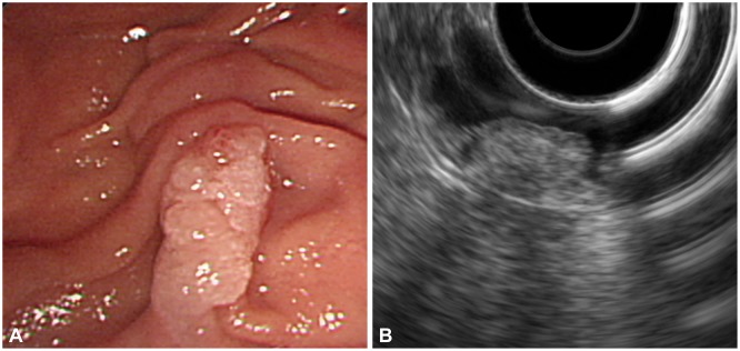
Fig. 1 Pre-procedural evaluation of ampullary adenoma. (A) Endoscopic exam demonstrates a pale, granular lesion on the major duodenal papilla. (B) Endoscopic ultrasonography with radial echoendoscope shows that the adenoma is limited to mucosa.
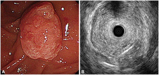
Fig. 2 Another case of preprocedural evaluation of ampullary adenoma. (A) Endoscopic exam shows a protuberant, hyperemic lesion at the major duodenal papilla. (B) Intraductal ultrasonography reveals that the adenoma is limited to mucosa and muscularis propria is intact (Kindly provided by Drs. Jong Ho Moon and Hyun Jong Choi from SoonchunHyang University Bucheon Hospital).
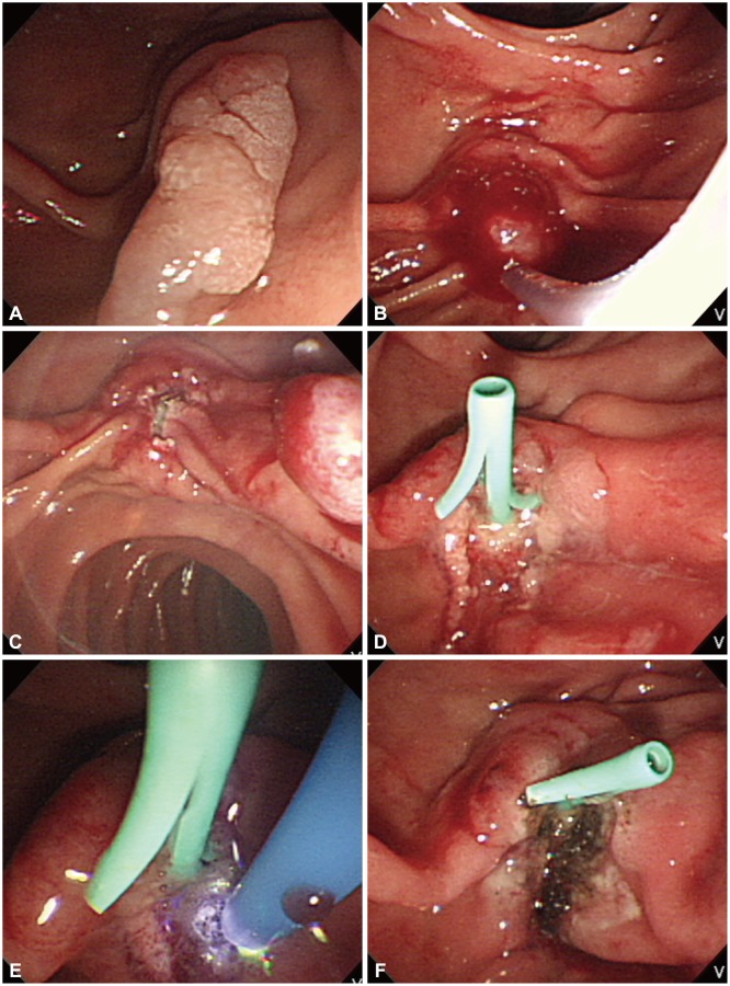
Fig. 3 Endoscopic papillectomy of ampullary adenoma. (A) A pale, elongated lesion is seen on the major duodenal papilla. (B) The adenoma is grasped with a standard polypectomy snare. (C) The tumor is removed en bloc after application of electrosurgical current. (D) A 5 Fr, 3 cm pancreatic stent is placed. (E) Hemostasis is achieved with argon plasma coagulation. (F) Postpapillectomy ulcer is clear without bleeding.
Reference
-
1. Suzuki K, Kantou U, Murakami Y. Two cases with ampullary cancer who underwent endosopic excision. Prog Dig Endosc. 1983; 23:236–239.2. Binmoeller KF, Boaventura S, Ramsperger K, Soehendra N. Endoscopic snare excision of benign adenomas of the papilla of Vater. Gastrointest Endosc. 1993; 39:127–131. PMID: 8495831.
Article3. Zádorová Z, Dvofák M, Hajer J. Endoscopic therapy of benign tumors of the papilla of Vater. Endoscopy. 2001; 33:345–347. PMID: 11315897.
Article4. Desilets DJ, Dy RM, Ku PM, et al. Endoscopic management of tumors of the major duodenal papilla: refined techniques to improve outcome and avoid complications. Gastrointest Endosc. 2001; 54:202–208. PMID: 11474391.
Article5. Norton ID, Gostout CJ, Baron TH, Geller A, Petersen BT, Wiersema MJ. Safety and outcome of endoscopic snare excision of the major duodenal papilla. Gastrointest Endosc. 2002; 56:239–243. PMID: 12145603.
Article6. Kahaleh M, Shami VM, Brock A, et al. Factors predictive of malignancy and endoscopic resectability in ampullary neoplasia. Am J Gastroenterol. 2004; 99:2335–2339. PMID: 15571579.
Article7. Catalano MF, Linder JD, Chak A, et al. Endoscopic management of adenoma of the major duodenal papilla. Gastrointest Endosc. 2004; 59:225–232. PMID: 14745396.
Article8. Cheng CL, Sherman S, Fogel EL, et al. Endoscopic snare papillectomy for tumors of the duodenal papillae. Gastrointest Endosc. 2004; 60:757–764. PMID: 15557951.
Article9. Bohnacker S, Seitz U, Nguyen D, et al. Endoscopic resection of benign tumors of the duodenal papilla without and with intraductal growth. Gastrointest Endosc. 2005; 62:551–560. PMID: 16185970.
Article10. Harewood GC, Pochron NL, Gostout CJ. Prospective, randomized, controlled trial of prophylactic pancreatic stent placement for endoscopic snare excision of the duodenal ampulla. Gastrointest Endosc. 2005; 62:367–370. PMID: 16111953.
Article11. Han J, Lee SK, Park DH, et al. Treatment outcome after endoscopic papillectomy of tumors of the major duodenal papilla. Korean J Gastroenterol. 2005; 46:110–119. PMID: 16118521.12. Jung MK, Cho CM, Park SY, et al. Endoscopic resection of ampullary neoplasms: a single-center experience. Surg Endosc. 2009; 23:2568–2574. PMID: 19360365.
Article13. Irani S, Arai A, Ayub K, et al. Papillectomy for ampullary neoplasm: results of a single referral center over a 10-year period. Gastrointest Endosc. 2009; 70:923–932. PMID: 19608181.
Article14. Boix J, Lorenzo-Zúñiga V, Moreno de Vega V, Domènech E, Gassull MA. Endoscopic resection of ampullary tumors: 12-year review of 21 cases. Surg Endosc. 2009; 23:45–49. PMID: 18398649.
Article15. Yamao T, Isomoto H, Kohno S, et al. Endoscopic snare papillectomy with biliary and pancreatic stent placement for tumors of the major duodenal papilla. Surg Endosc. 2010; 24:119–124. PMID: 19517183.
Article16. Nguyen N, Shah JN, Binmoeller KF. Outcomes of endoscopic papillectomy in elderly patients with ampullary adenoma or early carcinoma. Endoscopy. 2010; 42:975–977. PMID: 21072717.
Article17. Heinzow HS, Lenz P, Lenze F, Domagk D, Domschke W, Meister T. Feasibility of snare papillectomy in ampulla of Vater tumors: meta-analysis and study results from a tertiary referral center. Hepatogastroenterology. 2012; 59:332–335. PMID: 21940377.
Article18. Will U, Muller AK, Fueldner F, Wanzar I, Meyer F. Endoscopic papillectomy: data of a prospective observational study. World J Gastroenterol. 2013; 19:4316–4324. PMID: 23885142.
Article19. Ridtitid W, Tan D, Schmidt SE, et al. Endoscopic papillectomy: risk factors for incomplete resection and recurrence during long-term follow-up. Gastrointest Endosc. 2014; 79:289–296. PMID: 24094466.
Article20. Napoleon B, Gincul R, Ponchon T, et al. Endoscopic papillectomy for early ampullary tumors: long-term results from a large multicenter prospective study. Endoscopy. 2014; 46:127–134. PMID: 24477368.
Article21. Ismail S, Marianne U, Heikki J, Jorma H, Leena K. Endoscopic papillectomy, single-centre experience. Surg Endosc. 2014; 28:3234–3239. PMID: 24928230.
Article22. Moon JH, Choi HJ, Lee YN. Current status of endoscopic papillectomy for ampullary tumors. Gut Liver. 2014; 8:598–604. PMID: 25368746.
Article23. De Palma GD. Endoscopic papillectomy: indications, techniques, and results. World J Gastroenterol. 2014; 20:1537–1543. PMID: 24587629.24. El Hajj II, Coté GA. Endoscopic diagnosis and management of ampullary lesions. Gastrointest Endosc Clin N Am. 2013; 23:95–109. PMID: 23168121.
Article25. Patel R, Varadarajulu S, Wilcox CM. Endoscopic ampullectomy: techniques and outcomes. J Clin Gastroenterol. 2012; 46:8–15. PMID: 22064552.26. Hernandez LV, Catalano MF. Endoscopic papillectomy. Curr Opin Gastroenterol. 2008; 24:617–622. PMID: 19122504.
Article27. Han J, Kim MH. Endoscopic papillectomy for adenomas of the major duodenal papilla (with video). Gastrointest Endosc. 2006; 63:292–301. PMID: 16427938.
Article28. Standards of Practice Committee. Adler DG, Qureshi W, et al. The role of endoscopy in ampullary and duodenal adenomas. Gastrointest Endosc. 2006; 64:849–854. PMID: 17140885.
Article29. Bohnacker S, Soehendra N, Maguchi H, Chung JB, Howell DA. Endoscopic resection of benign tumors of the papilla of vater. Endoscopy. 2006; 38:521–525. PMID: 16767591.
Article30. Castillo C. Endoscopic ultrasound in the papilla and the periampullary region. World J Gastrointest Endosc. 2010; 2:278–287. PMID: 21160627.
Article31. Cannon ME, Carpenter SL, Elta GH, et al. EUS compared with CT, magnetic resonance imaging, and angiography and the influence of biliary stenting on staging accuracy of ampullary neoplasms. Gastrointest Endosc. 1999; 50:27–33. PMID: 10385718.
Article32. Skordilis P, Mouzas IA, Dimoulios PD, Alexandrakis G, Moschandrea J, Kouroumalis E. Is endosonography an effective method for detection and local staging of the ampullary carcinoma? A prospective study. BMC Surg. 2002; 2:1. PMID: 11914153.
Article33. Trikudanathan G, Njei B, Attam R, Arain M, Shaukat A. Staging accuracy of ampullary tumors by endoscopic ultrasound: meta-analysis and systematic review. Dig Endosc. 2014; 26:617–626. PMID: 24533918.
Article34. Chen CH, Tseng LJ, Yang CC, Yeh YH, Mo LR. The accuracy of endoscopic ultrasound, endoscopic retrograde cholangiopancreatography, computed tomography, and transabdominal ultrasound in the detection and staging of primary ampullary tumors. Hepatogastroenterology. 2001; 48:1750–1753. PMID: 11813616.35. Artifon EL, Couto D Jr, Sakai P, da Silveira EB. Prospective evaluation of EUS versus CT scan for staging of ampullary cancer. Gastrointest Endosc. 2009; 70:290–296. PMID: 19523619.
Article36. Chen CH, Tseng LJ, Yang CC, Yeh YH. Preoperative evaluation of periampullary tumors by endoscopic sonography, transabdominal sonography, and computed tomography. J Clin Ultrasound. 2001; 29:313–321. PMID: 11424095.
Article37. Manta R, Conigliaro R, Castellani D, et al. Linear endoscopic ultrasonography vs magnetic resonance imaging in ampullary tumors. World J Gastroenterol. 2010; 16:5592–5597. PMID: 21105192.38. Chen CH, Yang CC, Yeh YH, Chou DA, Nien CK. Reappraisal of endosonography of ampullary tumors: correlation with transabdominal sonography, CT, and MRI. J Clin Ultrasound. 2009; 37:18–25. PMID: 18726967.
Article39. Wee E, Lakhtakia S, Gupta R, et al. The diagnostic accuracy and strength of agreement between endoscopic ultrasound and histopathology in the staging of ampullary tumors. Indian J Gastroenterol. 2012; 31:324–332. PMID: 22996048.
Article40. Itoh A, Goto H, Naitoh Y, Hirooka Y, Furukawa T, Hayakawa T. Intraductal ultrasonography in diagnosing tumor extension of cancer of the papilla of Vater. Gastrointest Endosc. 1997; 45:251–260. PMID: 9087831.
Article41. Okano N, Igarashi Y, Hara S, et al. Endosonographic preoperative evaluation for tumors of the ampulla of vater using endoscopic ultrasonography and intraductal ultrasonography. Clin Endosc. 2014; 47:174–177. PMID: 24765600.
Article42. Menzel J, Hoepffner N, Sulkowski U, et al. Polypoid tumors of the major duodenal papilla: preoperative staging with intraductal US, EUS, and CT: a prospective, histopathologically controlled study. Gastrointest Endosc. 1999; 49(3 Pt 1):349–357. PMID: 10049419.43. Moon JH. Endoscopic diagnosis of ampullary tumors using conventional endoscopic ultrasonography and intraductal ultrasonography in the era of endoscopic papillectomy: advantages and limitations. Clin Endosc. 2014; 47:127–128. PMID: 24765593.
Article44. Singh P, Das A, Isenberg G, et al. Does prophylactic pancreatic stent placement reduce the risk of post-ERCP acute pancreatitis? A meta-analysis of controlled trials. Gastrointest Endosc. 2004; 60:544–550. PMID: 15472676.
Article45. Chini P, Draganov PV. Diagnosis and management of ampullary adenoma: the expanding role of endoscopy. World J Gastrointest Endosc. 2011; 3:241–247. PMID: 22195233.
Article46. Chang WI, Min YW, Yun HS, et al. Prophylactic pancreatic stent placement for endoscopic duodenal ampullectomy: a single-center retrospective study. Gut Liver. 2014; 8:306–312. PMID: 24827628.
Article47. Moon JH, Cha SW, Cho YD, et al. Wire-guided endoscopic snare papillectomy for tumors of the major duodenal papilla. Gastrointest Endosc. 2005; 61:461–466. PMID: 15758926.
Article48. Keenan J, Mallery S, Freeman ML. EUS rendezvous for pancreatic stent placement during endoscopic snare ampullectomy. Gastrointest Endosc. 2007; 66:850–853. PMID: 17719045.
Article49. Hwang JC, Kim JH, Lim SG, Yoo BM, Cho SW. Endoscopic resection of ampullary adenoma after a new insulated plastic pancreatic stent placement: a pilot study. J Gastroenterol Hepatol. 2010; 25:1381–1385. PMID: 20659227.
Article50. Nakahara K, Okuse C, Suetani K, et al. A novel endoscopic papillectomy after a pancreatic stent placement above the pancreatic duct orifice: inside pancreatic stenting papillectomy. J Clin Gastroenterol. 2014; 48:796–800. PMID: 24177378.
- Full Text Links
-
- Actions
-
Cited
- CITED
-
- Close
- Share
-
- Similar articles
-
- Endosonographic Preoperative Evaluation for Tumors of the Ampulla of Vater Using Endoscopic Ultrasonography and Intraductal Ultrasonography
- The Prevention of Pancreatitis after Endoscopic Papillectomy; Stent versus No Stent
- Role of transduodenal ampullectomy for tumors of the ampulla of Vater
- Unsolved problems in endoscopic papillectomy
- Endoscopic Management of Ampullary Tumors
