Ann Dermatol.
2009 May;21(2):182-184. 10.5021/ad.2009.21.2.182.
Mycosis Fungoides as an Ichthyosiform Eruption
- Affiliations
-
- 1Department of Dermatology, Chonbuk National University Medical School, Jeonju, Korea. dermayun@chonbuk. ac.kr
- 2Department of Plastic Surgery, Chonbuk National University Medical School, Jeonju, Korea.
- 3Department of Pathology, Samsung Medical Center, Sungkyunkwan University School of Medicine, Seoul, Korea.
- KMID: 2219392
- DOI: http://doi.org/10.5021/ad.2009.21.2.182
Abstract
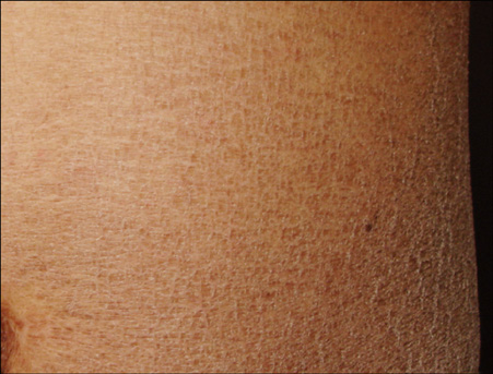
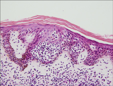
- Ichthyosiform eruption as a specific manifestation of mycosis fungoides is very rare and only a few such cases have currently been reported in the medical literature. A 63- year-old Korean man presented with a 4-year history of a pruritic ichthyotic eruption. There was no personal or family history of ichthyosis or atopy. The ichthyosiform skin changes involved the abdomen, arms, thighs and shins. The face, palms and soles were spared. There was no peripheral lymphadenopathy or organomegaly. The typical lesions of mycosis fungoides were not present. The results of the routine investigations were normal or negative. A skin biopsy specimen revealed the findings of early mycosis fungoides. He was successfully treated with photochemotherapy.
Keyword
Figure
Reference
-
1. Kazakov DV, Burg G, Kempf W. Clinicopathological spectrum of mycosis fungoides. J Eur Acad Dermatol Venereol. 2004. 18:397–415.
Article2. Bianchi L, Papoutsaki M, Orlandi A, Citarella L, Chimenti S. Ichthyosiform mycosis fungoides: a neoplastic acquired ichthyosis. Acta Derm Venereol. 2007. 87:82–83.
Article3. Sato M, Sohara M, Kitamura Y, Hatamochi A, Yamazaki S. Ichthyosiform mycosis fungoides: report of a case associated with IgA nephropathy. Dermatology. 2005. 210:324–328.
Article4. Hodak E, Amitay I, Feinmesser M, Aviram A, David M. Ichthyosiform mycosis fungoides: an atypical variant of cutaneous T-cell lymphoma. J Am Acad Dermatol. 2004. 50:368–374.
Article5. Eisman S, O'Toole EA, Jones A, Whittaker SJ. Granulomatous mycosis fungoides presenting as an acquired ichthyosis. Clin Exp Dermatol. 2003. 28:174–176.
Article6. Badawy E, D'Incan M, El Majjaoui S, Franck F, Fabricio L, Dereure O, et al. Ichthyosiform mycosis fungoides. Eur J Dermatol. 2002. 12:594–596.7. Marzano AV, Borghi A, Facchetti M, Alessi E. Ichthyosiform mycosis fungoides. Dermatology. 2002. 204:124–129.
Article8. Kutting B, Metze D, Luger TA, Bonsmann G. Mycosis fungoides presenting as an acquired ichthyosis. J Am Acad Dermatol. 1996. 34:887–889.
Article9. Signoretti S, Murphy M, Cangi MG, Puddu P, Kadin ME, Loda M. Detection of clonal T-cell receptor gamma gene rearrangements in paraffin-embedded tissue by polymerase chain reaction and nonradioactive single-strand conformational polymorphism analysis. Am J Pathol. 1999. 154:67–75.
Article10. Schwartz RA, Williams ML. Acquired ichthyosis: a marker for internal disease. Am Fam Physician. 1984. 29:181–184.


