Ann Dermatol.
2009 May;21(2):159-163. 10.5021/ad.2009.21.2.159.
Two Cases of Linear Alopecia on the Occipital Scalp
- Affiliations
-
- 1Department of Dermatology, Chonbuk National University Medical School, Jeonju, Korea. cwihm@chonbuk.ac.kr
- 2Department of Dermatology, Dankook University Hospital, Cheonan, Korea.
- KMID: 2219386
- DOI: http://doi.org/10.5021/ad.2009.21.2.159
Abstract
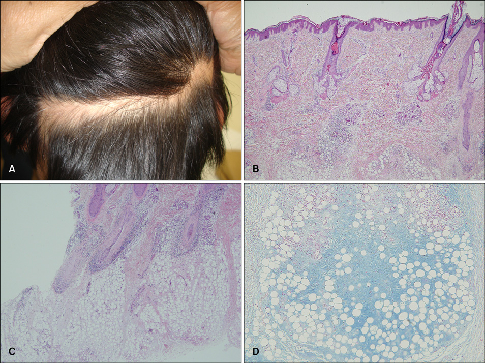
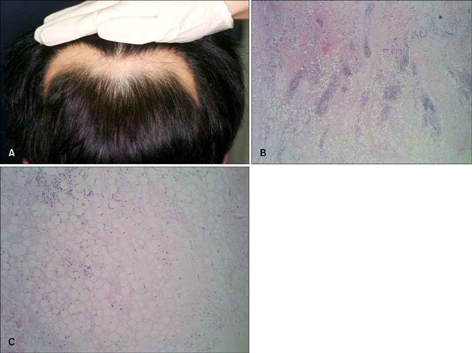
- Alopecia of a scalp shows various shapes and extents of hair loss, from a small round patch to polymorphous patches or total global alopecia. But alopecia of a linear shape is very rare. Only a few such cases have currently been reported in the medical literature. We recently had the chance to observe and treat two cases of linear alopecia that developed on the occipital scalp. The lesions themselves were like alopecia areata that shows a smooth bald area without any abnormality except the hair loss, but histopathologically, the lesions were compatible with lupus erythematosus profundus.
Keyword
MeSH Terms
Figure
Reference
-
1. Tagra S, Talwar AK, Walia RL. Lines of Blaschko. Indian J Dermatol Venereol Leprol. 2005. 71:57–59.
Article2. Nagai Y, Ishikawa O, Hattori T, Ogawa T. Linear lupus erythematosus profundus on the scalp following the lines of Blaschko. Eur J Dermatol. 2003. 13:294–296.3. Shin MK, Cho TH, Lew BL, Sim WY. A case of linear lupus erythematosus profundus on the scalp presenting as alopecia. Korean J Dermatol. 2007. 45:1280–1283.4. Jaworsky C. Elder DE, Elenitsas R, Johnson BL, Murphy GF, editors. Connective tissue diseases. Lever's histopathology of the skin. 2005. 9th ed. Philadelphia: Lippincott Williams & Wilkins;293–322.5. Kossard S. Lupus panniculitis clinically simulating alopecia areata. Australas J Dermatol. 2002. 43:221–223.
Article6. Werth VP, White WL, Sanchez MR, Franks AG. Incidence of alopecia areata in lupus erythematosus. Arch Dermatol. 1992. 128:368–371.
Article7. Constner MI, Sontheimer RD. Wolff K, Goldsmith LA, Katz SI, Gilchrest BA, Paller AS, Leffell DJ, editors. Lupus erythematosus. Fitzpatrick's dermatology in general medicine. 2008. 7th ed. New York: McGraw-Hill;1525–1526.8. Tada J, Arata J, Katayama H. Linear lupus erythematosus profundus in a child. J Am Acad Dermatol. 1991. 24:871–874.
Article9. Innocenzi D, Pranteda G, Giombini S, Silipo V, Bottoni U, Calvieri S. Linear lupus erythematosus profundus in an adolescent. Eur J Dermatol. 1997. 7:445–447.10. Tamada Y, Arisawa S, Ikeya T, Yokoi T, Hara K, Matsumoto Y. Linear lupus erythematosus profundus in a young man. Br J Dermatol. 1999. 140:177–178.
Article11. Choi WS, Kim JW, Park HS, Jang SJ, Choi JC. A case of linear lupus panniculitis in child. Korean J Dermatol. 2006. 44:1367–1369.12. Bolognia JL, Orlow SJ, Glick SA. Lines of Blaschko. J Am Acad Dermatol. 1994. 31:157–190.
Article
- Full Text Links
- Actions
-
Cited
- CITED
-
- Close
- Share
- Similar articles
-
- Histopathology of Alopecia Areata and Male Androgenetic Alopecia in Horizontal Sections of Scalp Biopsies in Koreans
- Two Cases of "Pinch Modification" of the Linear Advancement Flap (Peng Flap) in Repair of Round Defects on the Scalp
- Histopathologic findings of normal scalp and alopecia areata in transverse sections
- Congenital Triangular Alopecia
- Alopecia Associated with Underlying Congenital Melanocytic Nevus


