A Case of Multiple Eyelid Trichilemmoma Associated with Cowden Syndrome
- Affiliations
-
- 1Department of Ophthalmology, Yeungnam University College of Medicine, Daegu, Korea. sjh@med.yu.ac.kr
- KMID: 2217050
- DOI: http://doi.org/10.3341/jkos.2013.54.5.803
Abstract
- PURPOSE
To report a case of multiple eyelid trichilemmomas associated with Cowden syndrome.
CASE SUMMARY
A 27-year-old woman presented with multiple upper and lower eyelid skin masses that developed over several years. The masses were as large as whitish millet, and were around the upper and lower eyelid margin and the face. The patient had previously undergone subtotal thyroidectomy for a thyroid mass and a mass excision for extremity hemangioma. Excisional biopsy was performed for the diagnosis, and trichilemmoma was diagnosed based on histopathologic examination. Consequently, multiple trichilemmoma associated with Cowden syndrome was diagnosed, and breast evaluations for existence of further masses were recommended. On breast examinations, intraductal papilloma and fibroadenoma were detected.
CONCLUSIONS
The trichilemmoma was a hair-follicle benign tumor that also appeared on the skin around the eyelid. If multiple trichilemmoma is diagnosed, an association with Cowden syndrome should be considered as well as presence of masses in other organs.
Keyword
MeSH Terms
Figure
-
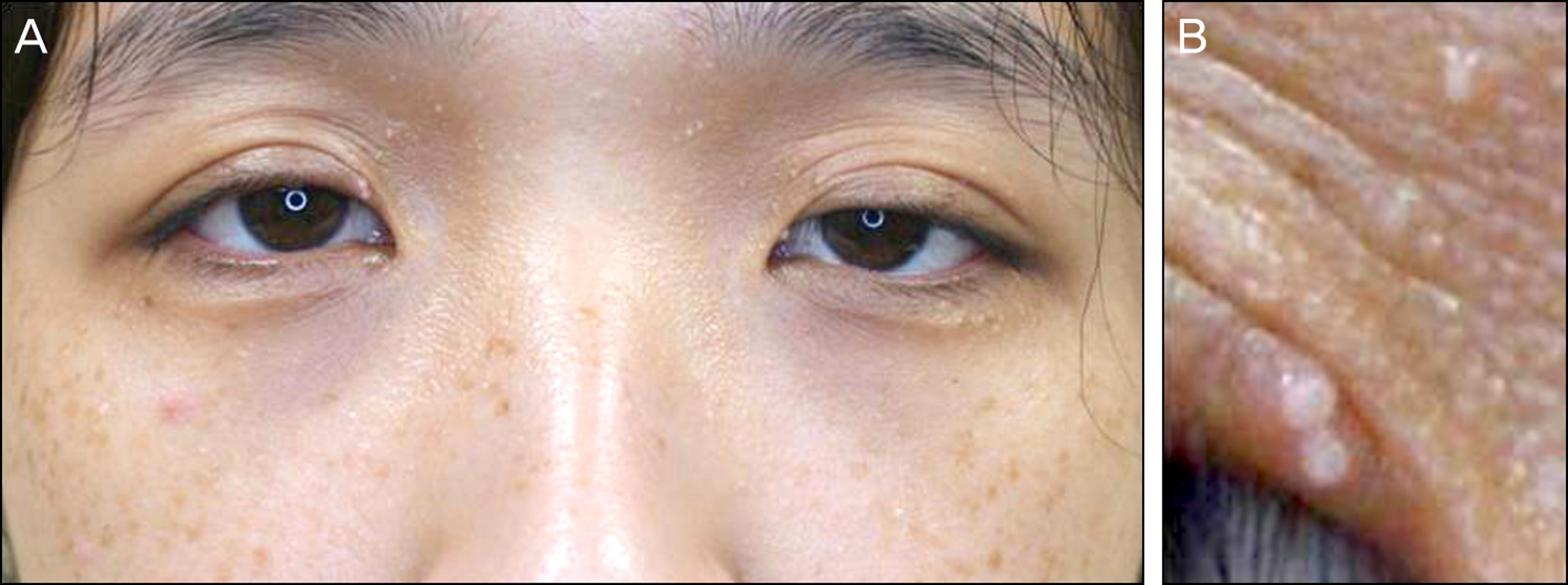
Figure 1. External appearance of the patient. (A) She pre-sented with multiple whitish masses on her upper and lower eyelids and face. (B) Magnified image of the right upper medial canthus. A milium-sized whit-ish mass is seen.

Figure 2. Surgical biopsy of the eyelid mass. (A) The tumor showed lobular, follicle-centered proliferation with overlying keratosis (arrow head; hematoxylin and eosin stain, ×40) (B) The tumor was composed of bland, amitotic, uniform polyhedral cells with clear glycogenated cytoplasm (small arrow), surrounded by cuffs of basement membrane material. (hematoxylin and eosin stain, ×100) (C) All tumor cells demonstrate basal cell peripheral nuclear palisading pattern in the cellular lobules (big arrow; hematoxylin and eosin stain, ×100) The tumor was diagnosed as trichilemmoma.

Figure 3. Mammogram (A) and specimen (B) of the right breast. (A) Small oval isoechogenic nodules in cystic echoes and ductal dilatation were seen at the subareolar area (suspected intraductal papilloma). (B) Specimen obtained from the right breast mass after surgical excision. The cut surface shows a dilated duct with solid mural nodule. The tumor was finally di-agnosed as fibroadenoma with a complex sclerosing lesion and intraductal papilloma with florid ductal hyperplasia.
Reference
-
References
1. Headington JT, French AJ. Primary neoplasms of the hair follicle. Histogenesis and classification. Arch Dermatol. 1962; 86:430–41.
Article2. Brownstein MH, Shapiro L. Trichilemmoma. Analysis of 40 new cases. Arch Dermatol. 1973; 107:866–9.
Article3. Brownstein MH, Wolf M, Bikowski JB. Cowden's disease: a cuta-neous marker of breast cancer. Cancer. 1978; 41:2393–8.4. Hong WK, Song HJ, Lee HS, et al. A case of cowden syndrome. Korean J Dermatol. 2007; 45:829–31.5. Eng C. Will the real Cowden syndrome please stand up: revised di-agnostic criteria. J Med Genet. 2000; 37:828–30.
Article6. Starink TM. Cowden's disease: analysis of fourteen new cases. J Am Acad Dermatol. 1984; 11:1127–41.
Article7. Lloyd KM 2nd, Dennis M. Cowden's disease. A possible new symptom complex with multiple system involvement. Ann Intern Med. 1963; 58:136–42.8. Rustad CF, Bjørnslet M, Heimdal KR, et al. Germline PTEN muta-tions are rare and highly penetrant. Hered Cancer Clin Pract. 2006; 4:177–85.
Article9. Al-Zaid T, Ditelberg JS, Prieto VG, et al. Trichilemmomas show loss of PTEN in cowden syndrome but only rarely in sporadic tumors. J Cutan Pathol. 2012; 39:493–9.
Article10. Masmoudi A, Chermi ZM, Marrekchi S, et al. Cowden syndrome. J Dermatol Case Rep. 2011; 5:8–13.
Article11. Ha M, Chung JW, Hahm KB, et al. A case of Cowden syndrome di-agnosed from multiple gastric polyposis. World J Gastroenterol. 2012; 18:861–4.
Article12. Schweitzer S, Hogge JP, Grimes M, et al. Cowden disease: a cuta-neous marker for increased risk of breast cancer. AJR Am J Roentgenol. 1999; 172:349–51.
Article13. Hildenbrand C, Burgdorf WH, Lautenschlager S. Cowden syn-drome-diagnostic skin signs. Dermatology. 2001; 202:362–6.14. Ravi Prakash SM, Suma GN, Goel S. Cowden syndrome. Indian J Dent Res. 2010; 21:439–42.
Article15. Jung MH. Breast cancer in cowden syndrome : a case report. J Korean Soc Radiol. 2009; 60:279–82.16. Rohwedder A, Keminer O, Hendricks C, Schaller J. Detection of HPV DNA in trichilemmomas by polymerase chain reaction. J Med Virol. 1997; 51:119–25.
Article17. Schaller J, Rohwedder A, Burgdorf WH, et al. Identification of hu-man papillomavirus DNA in cutaneous lesions of cowden syndrome. Dermatology. 2003; 207:134–40.
Article
