Location of the Tube Tip in the Anterior Chamber and Change in Corneal Endothelium after Ahmed Valve Implantation
- Affiliations
-
- 1Jongro Kong Eye Clinic, Seoul, Korea.
- 2Department of Ophthalmology, Seoul National University College of Medicine, Seoul, Korea. kihopark@snu.ac.kr
- 3Department of Ophthalmology, Seoul National University Bundang Hospital, Seongnam, Korea.
- 4Department of Ophthalmology, Seoul National University Hospital, Seoul, Korea.
- KMID: 2216768
- DOI: http://doi.org/10.3341/jkos.2013.54.3.469
Abstract
- PURPOSE
To evaluate the impact of location of a silicone tube tip in the anterior chamber on corneal endothelium after Ahmed glaucoma valve implantation.
METHODS
We measured the distance from the tip of a silicone tube in the anterior chamber to the posterior surface of the cornea by anterior segment optical coherence tomography (OCT) in 24 eyes of 21 patients who underwent Ahmed glaucoma valve implantation. All surgeries were performed by a single surgeon. The corneal endothelial cells of central, superior, superotemporal, and superonasal area of the cornea were examined by specular microscope before and after surgery.
RESULTS
The distance from the tip of the silicone tube in the anterior chamber to the posterior surface of the cornea and the corneal endothelium were measured at 19.2 +/- 11.8 months (2.8-41.2 months) after surgery. At the central corneal area, a statistically significant decrease in the number of corneal endothelial cells (2278 +/- 565/mm2 vs. 2177 +/- 529/mm2, p = 0.043) after surgery was observed, but no relationship was found between the amount of decrease and distance from the tip to the posterior surface of the cornea. At the superotemporal corneal area where the tip of the silicone tube was located in the anterior chamber, there was more significant loss of corneal endothelial cells than in the other areas after surgery (p = 0.006). Moreover, the amount of endothelial cell loss at the superotemporal area was negatively correlated to the distance from the tip to the posterior surface of the cornea (partial correlation coefficient by time -0.558, p = 0.031).
CONCLUSIONS
To minimize the loss of corneal endothelial cells after Ahmed glaucoma valve implantation, ensuring a sufficient distance from the tip of the silicone tube to the posterior surface of the cornea is important.
Keyword
MeSH Terms
Figure
-
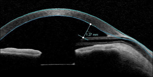
Figure 1. Visante OCT single scan along long axis of the silicone tube of Ahmed glaucoma valve showed the position and patency of the silicone tube in the anterior chamber. The distance from the tip of silicone tube to the posterior surface of cornea was measured to be 1.27 mm.
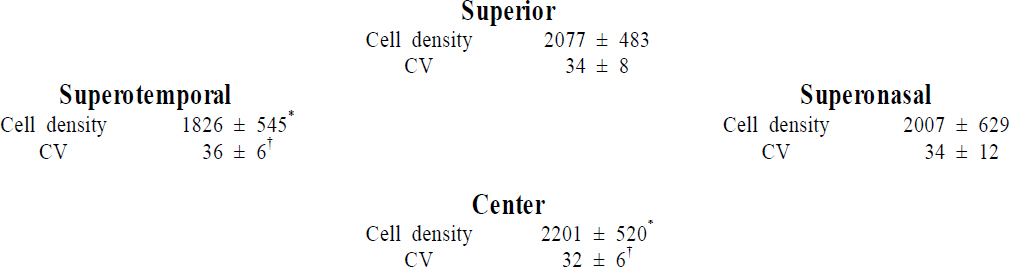
Figure 2. Topographical corneal endothelial cell density and coefficient of variation of cell area (CV) after Ahmed glaucoma valve implantation. The cell density and CV at superotemporal corneal area where the tip of the silicone tube was located in the anterior chamber got worse, compared to those at central corneal area.Values are presented as mean ± SD, Cell density (cells/mm2), Coefficient of variation of cell area (CV). *p = 0.006, W ilcoxon signed ranks test; †p = 0.013, W ilcoxon signed ranks test.
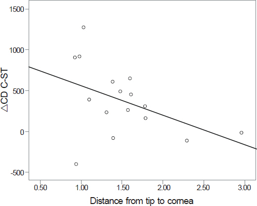
Figure 3. The difference of corneal endothelial cell density between central and superotemporal corneal area (ΔCD C-ST) was negatively correlated to the distance from the tip of the silicone tube in the anterior chamber to the posterior surface of the cornea (partial correlation coefficient adjusted by time -0.558, p = 0.031).
Cited by 2 articles
-
Corneal Endothelial Cell Loss after Tube Shunt Surgery in Fuch's Heterochromic Iridocyclitis
Jin Ah Lee, Yang Kyung Cho, Tae Yoon La, Jin A Choi
J Korean Ophthalmol Soc. 2015;56(4):643-649. doi: 10.3341/jkos.2015.56.4.643.Risk Factors for Cataract Formation after Implantable Collamer Lens Implantation: Over a Mean 7.5-Year Follow-Up Period
Damho Lee, Ju Yong Seok, Hak Su Kyung, Joon Mo Kim
J Korean Ophthalmol Soc. 2015;56(6):835-846. doi: 10.3341/jkos.2015.56.6.835.
Reference
-
References
1. Gedde SJ, Schiffman JC, Feuer WJ, et al. Treatment outcomes in the Tube Versus Trabeculectomy (TVT) study after five years of follow-up. Am J Ophthalmol. 2012; 153:789–803.
Article2. Taglia DP, Perkins TW, Gangnon R, et al. Comparison of the Ahmed Glaucoma Valve, the Krupin Eye Valve with Disk, and the double-plate Molteno implant. J Glaucoma. 2002; 11:347–53.
Article3. Hong CH, Arosemena A, Zurakowski D, Ayyala RS. Glaucoma drainage devices: a systematic literature review and current controversies. Surv Ophthalmol. 2005; 50:48–60.
Article4. Kim JH, Kim CS. The change in corneal endothelial cells after Ahmed Glaucoma Valve implantation. J Korean Ophthalmol Soc. 2006; 47:1972–80.5. Wells AP, Barton K, Konstas AG. Corneal edema after aqueous drainage device implantation. J Glaucoma. 2007; 16:388–90.
Article6. McDermott ML, Swendris RP, Shin DH, et al. Corneal endothelial cell counts after Molteno implantation. Am J Ophthalmol. 1993; 115:93–6.
Article7. Fiore PM, Richter CU, Arzeno G, et al. The effect of anterior chamber depth on endothelial cell count after filtration surgery. Arch Ophthalmol. 1989; 107:1609–11.
Article8. Sarodia U, Sharkawi E, Hau S, Barton K. Visualization of aqueous shunt position and patency using anterior segment optical coherence tomography. Am J Ophthalmol. 2007; 143:1054–6.
Article9. Lee JJ, Park KH, Kim DM, Kim TW. Clinical outcomes of Ahmed glaucoma valve implantation using tube ligation and removable external stents. Korean J Ophthalmol. 2009; 23:86–92.
Article10. Catoira-Boyle YPM, WuDunn D, Cantor L. Postoperative complications. In : Shaarawy TM, Sherwood MB, Hitchings RA, Crowston JG, editors. Glaucoma Volume 2: Surgical Management. Saunders;2009. v. 2. chap. 103.11. Byun YS, Lee NY, Park CK. Risk factors of implant exposure outside the conjunctiva after Ahmed glaucoma valve implantation. Jpn J Ophthalmol. 2009; 53:114–9.
Article12. Kim J, Lee J, Kee C. Tissue incarceration after Ahmed valve implantation. J Korean Ophthalmol Soc. 2012; 53:1053–6.
Article13. Lee EK, Yun YJ, Lee JE, et al. Changes in corneal endothelial cells after Ahmed glaucoma valve implantation: 2-year follow-up. Am J Ophthalmol. 2009; 148:361–7.
Article14. Mendrinos E, Dosso A, Sommerhalder J, Shaarawy T. Coupling of HRT II and AS-OCT to evaluate corneal endothelial cell loss and in vivo visualization of the Ahmed glaucoma valve implant. Eye (Lond). 2009; 23:1836–44.
Article15. Lopilly Park HY, Jung KI, Park CK. Serial intracameral visualization of the Ahmed glaucoma valve tube by anterior segment optical coherence tomography. Eye (Lond). 2012; 26:1256–62.
Article16. Ramulu PY, Corcoran KJ, Corcoran SL, Robin AL. Utilization of various glaucoma surgeries and procedures in Medicare beneficiaries from 1995 to 2004. Ophthalmology. 2007; 114:2265–70.
Article17. Chen PP, Yamamoto T, Sawada A, et al. Use of antifibrosis agents and glaucoma drainage devices in the American and Japanese Glaucoma Societies. J Glaucoma. 1997; 6:192–6.
Article18. Joshi AB, Parrish RK 2nd, Feuer WF. 2002 Survey of the American Glaucoma Society: practice preferences for glaucoma surgery and antifibrotic use. J Glaucoma. 2005; 14:172–4.
- Full Text Links
-
- Actions
-
Cited
- CITED
-
- Close
- Share
-
- Similar articles
-
- Two Cases of Malignant Glaucoma after Ahmed Valve Implantation
- The Effect of Flat Anterior Chamber on the Success of Ahmed Valve Implantation
- A Case of Ahmed Glaucoma Valve Implantation without Removal of the Anterior Chamber Lens
- The Change in Corneal Endothelial Cells after Ahmed Glaucoma Valve Implantation
- The Effect of a 5-0 Prolene Ligation Around the Ahmed Glaucoma Valve Tube on the Postoperative Hypotony
