Successful Treatment of Bilateral Conjunctival-Corneal Intraepithelial Neoplasia: Case Report and Review of the Literature
- Affiliations
-
- 1Department of Ophthalmology, Inje University Ilsan Paik Hospital, Inje University College of Medicine, Goyang, Korea. jhk90924@hanmail.net
- KMID: 2216675
- DOI: http://doi.org/10.3341/jkos.2013.54.2.357
Abstract
- PURPOSE
We report a case of successfully treating bilateral conjunctival-corneal intraepithelial neoplasia (CCIN) with surgical excision and adjunctive cryotherapy.
CASE SUMMARY
A 74-year-old male visited our clinic for bilateral foreign body sensation and decreased visual acuity. His initial best corrected visual acuity was 20/50 in the right eye and 20/30 in the left eye. The right eye showed a 9 mm x 11 mm sized, gray-opaque limbal lesion from approximately the 7-o'clock position to the 11-o'clock position with spreading onto the cornea and conjunctiva. Biomicroscopy revealed a 6 mm x 7.5 mm sized minimally elevated, opaque lesion from the 3-o'clock to the 5-o'clock position extending to the central cornea in the left eye. The corneal lesion was well demarcated, opaque, and minimally elevated with bilateral focal pigmentation. Conjunctival lesions were finely vascularized and slightly elevated with melanocytic pigmentation. An excisional biopsy was performed to confirm the diagnosis and for therapeutic purposes, followed by an adjunctive cryotherapy. Postoperative corrected visual acuity improved up to 20/25 bilaterally and the patient had no recurrence 8 months after surgery.
CONCLUSIONS
Bilateral conjunctival-corneal intraepithelial neoplasia is a rare condition. We report successful treatment and control of recurrence in a patient with bilateral conjunctival-corneal intraepithelial neoplasia using conventional surgical excision and adjuvant cryotherapy rather than topical chemotherapy.
MeSH Terms
Figure
-
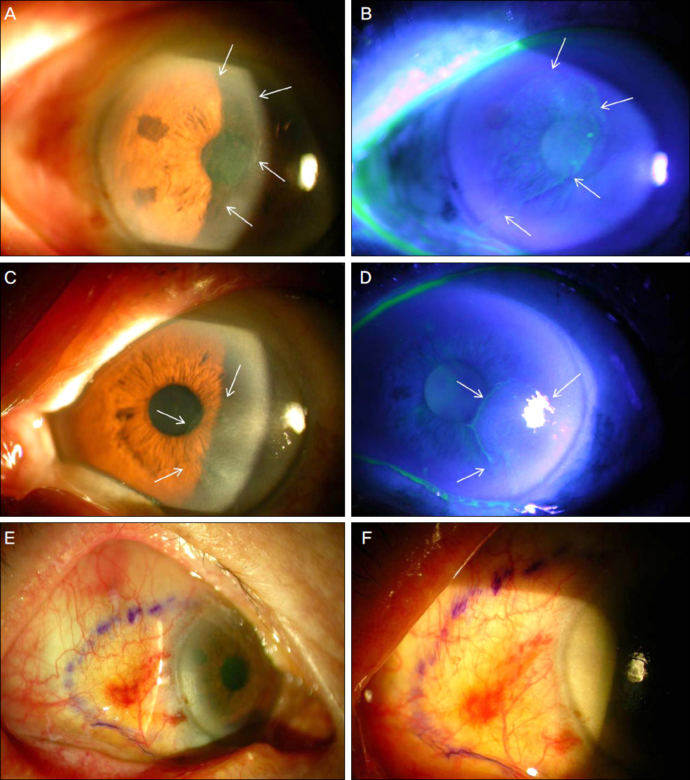
Figure 1. (A, B) Anterior segment photograph of right eye before surgery. Well demarcated the opaque lesion covering half of the corneal surface and extensively involving the visual axis. Note the hazy corneal component (arrows). (C, D) External photograph of the left eye showing a limbal to corneal gray and opaque lesion extending on to the visual axis from 3 to 5 O' clock position. (E, F) Relatively well demarcated yellowish elevated conjunctival lesion is seen within the violet marks.
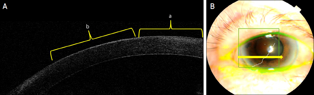
Figure 2. (A) Anterior segment OCT image of the cornea between the normal cornea (a) and CIN lesion (b). Slightly thickened epithelium (b) with hyperreflectivity is noticed compared with that of the normal corneal epithelium (a). (B) External photography showing corneal lesion of CCIN on the right eye. The arrow represents the direction of the OCT scan.
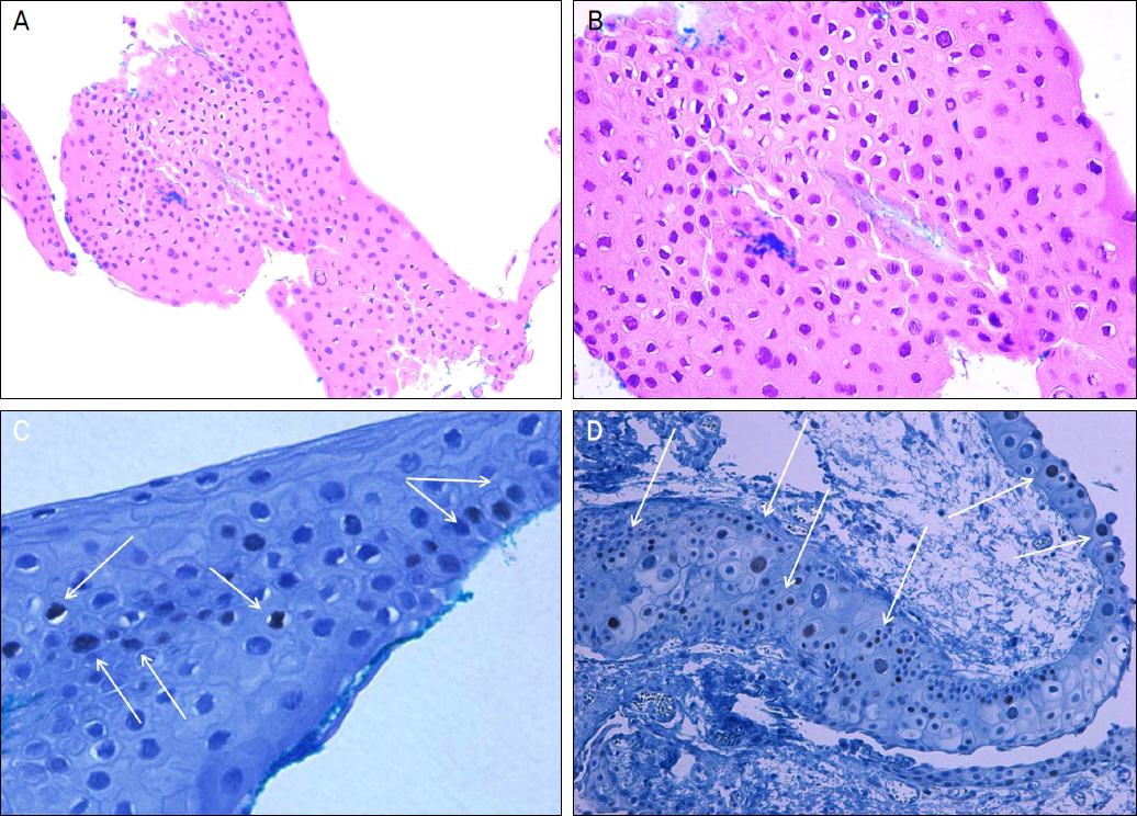
Figure 3. (A) Histopathologic photograph of excised tissue from the cornea showing thickened epithelium with scattered dyskeratotic cells and loss of polarity in the corneal epithelium (H-E stain, ×100). (B) Individual atypical cells with increased mitotic figures, increased nucleocytoplasmic ratio within the epithelium are seen (H-E stain, ×400). (C) Immunohistochemical staining of excised corneal epithelium showing moderate dysplasia and scattered brown nuclear staining (arrows) conferred to the basal and parabasal layers (p53, ×400). (D) The excised conjunctival tissue shows loss of polarity and scattered dys-plastic cells with p53 positivity (arrows) within the epithelium (p53, ×200).
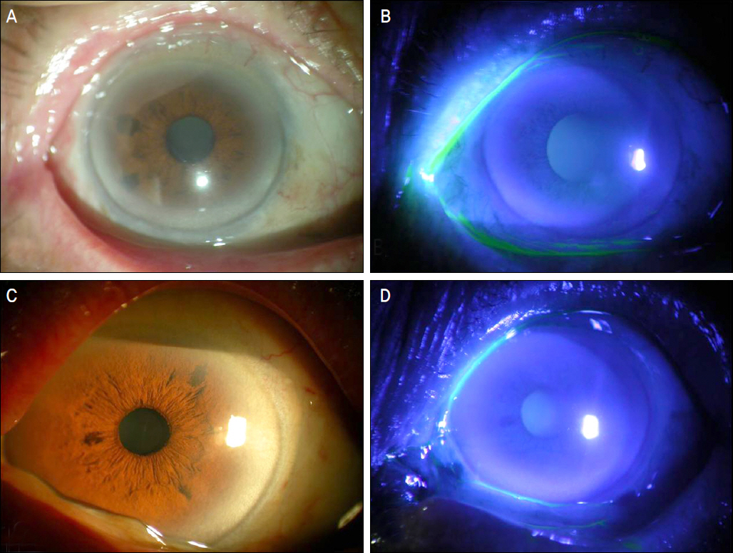
Figure 4. Anterior segment photograph of the right eye (A, B) and left eye (C, D) after 8 months of surgery exhibiting complete remission of conjunctival and corneal intraepithelial neoplasia.
Reference
-
References
1. Erie JC, Campbell RJ, Liesegang TJ. Conjunctival and corneal intraepithelial and invasive neoplasia. Ophthalmology. 1986; 93:176–83.
Article2. Waring GO 3rd, Roth AM, Elkins MB. Clinical and pathologic description of 17 cases of corneal intraepithelial neoplasia. AM J Ophthalmol. 1984; 97:547–59.
Article3. Basti S, Macsai MS. Ocular surface squamous neoplasia: a review. Cornea. 2003; 22:687–704.4. Waddell KM, Lewallen S, Lucas SB, et al. Carcinoma of the conjunctiva and HIV infection in Uganda and Malawi. Br J Ophthalmol. 1996; 80:503–8.
Article5. Lee Ga, Hirst LW. Retrospective study of ocular surface squamous neoplasia. Aust N Z J Ophthalmol. 1997; 25:269–76.
Article6. Cervantes G, Rodríguez AA Jr, Leal AG. Squamous cell carcinoma of the conjunctiva: clinicopathological features in 287 cases. Can J Ophthalmol. 2002; 37:14–9.
Article7. Schechter BA, Koreishi AF, Karp CL, Feuer W. Long-term follow-up of conjunctival and corneal intraepithelial neoplasia treated with topical interferon alfa-2b. Ophthalmology. 2008; 115:1291–6.
Article8. Krachmer, Mannis, Holland . Cornea. 2nd ed.Philadelphia: Elsevier Mosby;v. 2:2005. p. 1773–80.9. Frucht-Pery J, Rozenman Y. Mitomycin C therapy for corneal intraepithelial neoplasia. Am J Ophthalmol. 1994; 117:164–8.
Article10. Yeatts RP, Ford JG, Stanton CA, Reed JW. Topical 5-fluorouracil in treating epithelial neoplasia of the conjunctiva and cornea. Ophthalmology. 1995; 102:1338–44.
Article11. Park HJ, Lee JE, Lee JS, Park DY. Treatment of conjunctival- cor-neal intraepithelial neoplasia with topical mitomycin C. J Korean Ophthalmol Soc. 2003; 44:1924–30.12. Kim JW, Shin DE. A case of conjunctival carcinoma in situ. J Korean Ophthalmol Soc. 1986; 27:943–7.13. Lee JS, On KK, Kim JD. A case of corneal squamous cell carcinoma in situ. J Korean Ophthalmol Soc. 1994; 35:206–9.14. Lee DH, Jang JH, Oh JY, Kim JS. A case of conjunctival intraepithelial neoplasia (CIN) misdiagnosed as atypical pterygium. J Korean Ophthalmol Soc. 2000; 41:2750–4.15. Kim JH, Kang HS, Kim IC. The effect of topical mitomycin C after excisional biopsy in conjunctival-corneal intraepithelial neoplasia: two cases. J Korean Ophthalmol Soc. 2001; 42:1102–10.16. Cheon HC, Lee DY, Park WC, Ahn HB. Ocular surface squamous neoplasia. J Korean Ophthalmol Soc. 2006; 47:1920–8.17. Sun EC, Fears TR, Goedert JJ. Epidemiology of squamous cell conjunctival cancer. Cancer Epidemiol Biomarkers Prev. 1997; 6:73–7.18. Newton R, Ferlay J, Reeves G, et al. Effect of ambient solar ultra-violet radiation on incidence of squamous-cell carcinoma of the eye. Lancet. 1996; 347:1450–1.
Article19. Rundle P, Mudhar HS, Rennie I. Conjunctival intra-epithelial neoplasia occurring in young patients with asthma. Eye. 2010; 24:1182–5.
Article20. Odrich MG, Jakobiec FA, Lancaster WD, et al. A spectrum of bilateral squamous conjunctival tumors associated with human papillomavirus type 16. Ophthalmology. 1991; 98:628–35.
Article21. Tritten JJ, Beati D, Sahli R, Uffer S. [Bilateral conjunctivo-palpebral tumor in an immunocompetent man cause by human papilloma virus]. Klin Monbl Augenheilkd. 1994; 204:453–5.22. Gericke A, Pitz S, Strempel I, Sekundo W. [Bilateral ocular surface squamous neoplasia and neurodermatitis. Two cases with different courses]. Ophthalmologe. 2008; 105:1142–5.23. Guthoff R, Marx A, Stroebel P. No evidence for a pathogenic role of human papillomavirus infection in ocular surface squamous neoplasia in Germany. Curr Eye Res. 2009; 34:666–71.
Article24. McDonnell JM, McDonnell PJ, Sun YY. Human papillomavirus DNA in tissues and ocular surface swabs of patients with conjunctival epithelial neoplasia. Invest Ophthalmol Vis Sci. 1992; 33:184–9.25. Mahomed A, Chetty R. Human immunodeficiency virus infection, Bcl-2, p53 protein, and Ki-67 analysis in ocular surface squamous neoplasia. Arch Ophthalmol. 2002; 120:554–8.
Article26. Peksayar G, Altan-Yaycioglu R, Onal S. Excision and cryosurgery in the treatment of conjunctival malignant epithelial tumours. Eye. 2003; 17:228–32.
Article27. Rozenman Y, Frucht-Pery J. Treatment of conjunctival intraepithelial neoplasia with topical drops of mitomycin C. Cornea. 2000; 19:1–6.
Article28. Chen HC, Chang SW, Huang SF. Adjunctive treatment with inter-ferona alpha-2b may decrease the risk of papilloma-associated conjunctival intraepithelial neoplasm recurrence. Cornea. 2004; 23:726–9.
- Full Text Links
-
- Actions
-
Cited
- CITED
-
- Close
- Share
-
- Similar articles
-
- The Effect of Topical Mitomycin C after Excisional Biopsy in Conjunctival-corneal Intraepithelial Neoplasia: Two cases
- Treatment of Conjunctival-Corneal Intraepithelial Neoplasia with Topical Mitomycin C
- High-Grade Prostatic Intraepithelial Neoplasia
- Amniotic Membrane Transplantation for the Conjunctival Necrosis after Scleral Gra t in the Enucleated Eye: 1 Case Report
- Temporary Conjunctival Flap for the Treatment of Infectious Corneal Ulcer
