J Korean Ophthalmol Soc.
2013 Feb;54(2):289-295. 10.3341/jkos.2013.54.2.289.
Age-Related Differences of Spectral-Domain Optical Coherence Tomography Data in Koreans
- Affiliations
-
- 1Department of Ophthalmology, Dong-A University College of Medicine, Busan, Korea. shrho@donga.ac.kr
- KMID: 2216665
- DOI: http://doi.org/10.3341/jkos.2013.54.2.289
Abstract
- PURPOSE
We evaluated the thickness of RNFL and optic nerve head parameters with age in normal eyes using Spectral-Domain Optical Coherence Tomography (SD-OCT).
METHODS
A total of 128 normal Korean volunteers in different age groups were recruited (age range, 20-70 years).
RESULTS
A significant negative correlation in average RNFL thickness with increasing age was found. The inferior areas (130.31 +/- 3.33 micrometer) were significantly thicker than other areas (superior area 119.05 +/- 2.12 micrometer, nasal area 71.80 +/- 0.57 micrometer, temporal area 77.72 +/- 0.16 micrometer). The average C/D ratios (mean 0.38 +/- 0.14) and vertical C/D ratios (mean 0.35 +/- 0.11) both showed significant increases with age, and the vertical C/D ratio correlation coefficient was higher (average C/D ratio r = 0.249, vertical C/D ratio r = 0.537). However, rim area, disc area, and cup volume were not correlated with age.
CONCLUSIONS
From these findings, we conclude that, in normal Koreans, the mean RNFL thickness decreases and the C/D ratio increases with age, with the increase in the vertical C/D ratio being greater.
Keyword
Figure
-
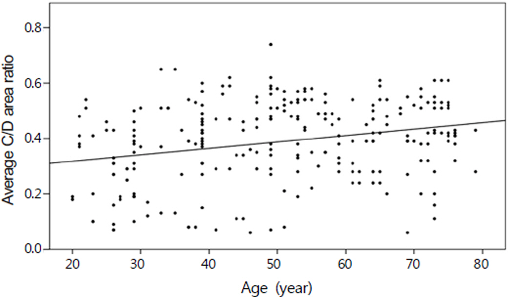
Figure 1. The relationship between average cup/disc (C/D) area ratio and age. Average C/D area ratio increases with age significantly (p < 0.05).
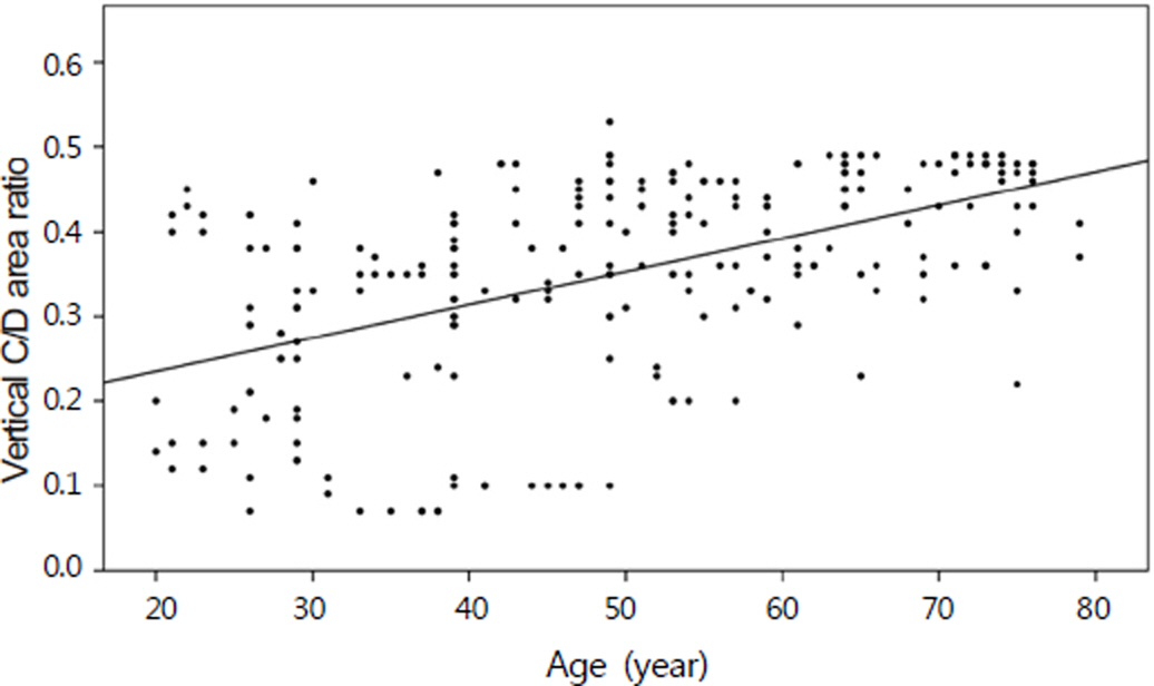
Figure 2. The relationship between vertical cup/disc (C/D) area ratio and age. Vertical C/D ratio increases with age significantly (p < 0.05).
Reference
-
References
1. Sugiyama K, Tomita G, Kitazawa Y, et al. The associations of optic disc hemorrhage with retinal nerve fiber layer defect and peripapillary atrophy in normal-tension glaucoma. Ophthalmology. 1997; 104:1926–33.
Article2. Tezel G, Trinkaus K, Wax MB. Alterations in the morphology of lamina cribrosa pores in glaucomatous eyes. Br J Ophthalmol. 2004; 88:251–6.
Article3. Quigley HA, Katz J, Derick RJ, et al. An evaluation of optic disc and nerve fiber layer examinations in monitoring progression of early glaucoma damage. Ophthalmology. 1992; 99:19–28.
Article4. Sommer A, Katz J, Quigley HA, et al. Clinically detectable nerve fiber atrophy precedes the onset of glaucomatous field loss. Arch Ophthalmol. 1991; 109:77–83.
Article5. Medeiros FA, Zangwill LM, Bowd C, Weinreb RN. Comparison of the GDx VCC scanning laser polarimeter, HRT II confocal scanning laser ophthalmoscope, and stratus OCT optical coherence tomograph for the detection of glaucoma. Arch Ophthalmol. 2004; 122:827–37.6. Zangwill LM, Bowd C, Berry CC, et al. Discriminating between normal and glaucomatous eyes using the Heidelberg Retina Tomograph, GDx Nerve Fiber Analyzer, and Optical Coherence Tomograph. Arch Ophthalmol. 2001; 119:985–93.
Article7. Bowd C, Weinreb RN, Williams JM, Zangwill LM. The retinal nerve fiber layer thickness in ocular hypertensive, normal, and glaucomatous eyes with optical coherence tomography. Arch Ophthalmol. 2000; 118:22–6.
Article8. Hoh ST, Greenfield DS, Mistlberger A, et al. Optical coherence tomography and scanning laser polarimetry in normal, ocular hyper-tensive, and glaucomatous eyes. Am J Ophthalmol. 2000; 129:129–35.
Article9. Hee MR, Izatt JA, Swanson EA, et al. Optical coherence tomog–raphy of the human retina. Arch Ophthalmol. 1995; 113:325–32.
Article10. Blumenthal EZ, Williams JM, Weinreb RN, et al. Reproducibility of nerve fiber layer thickness measurements by use of optical coherence tomography. Ophthalmology. 2000; 107:2278–82.11. Lalezary M, Medeiros FA, Weinreb RN, et al. Baseline optical coherence tomography predicts the development of glaucomatous change in glaucoma suspects. Am J Ophthalmol. 2006; 142:576–82.
Article12. Leung CK, Chan WM, Yung WH, et al. Comparison of macular and peripapillary measurements for the detection of glaucoma: an optical coherence tomography study. Ophthalmology. 2005; 112:391–400.13. Koizumi H, Spaide RF, Fisher YL, et al. Three-dimensional evaluation of vitreomacular traction and epiretinal membrane using spectraldomain optical coherence tomography. Am J Ophthalmol. 2008; 145:509–17.
Article14. Moon SW, Kim ES, Kim YG, et al. The comparison of macular thickness measurements and repeatabilities between time domain and spectral domain OCT. J Korean Ophthalmol Soc. 2009; 50:1050–9.
Article15. Lee JY, Hwang YH, Lee SM, Kim YY. Age and retinal nerve fiber layer thickness measured by spectral domain optical coherence tomography. Korean J Ophthalmol. 2012; 26:163–8.
Article16. Zeyen TG, Caprioli J. Progression of disc and field damage in early glaucoma. Arch Ophthalmol. 1993; 111:62–5.
Article17. Quigley HA, Addicks EM. Quantitiative studies of retinal nerve fiber layer defects. Arch Ophthalmol. 1982; 100:807–14.18. Hoyt WF, Frisén L, Newman NH. Fundoscopy of nerve fiber layer defects in glaucoma. Invest Ophthalmol. 1973; 12:814–29.19. Fercher AF, Hitzenberger CK, Drexler W, et al. In vivo optical coherence tomography. Am J Ophthalmol. 1993; 116:113–4.
Article20. Swanson EA, Izatt JA, Hee MR, et al. In vivo retinal imaging by optical coherence tomography. Opt Lett. 1993; 18:1864–6.
Article21. Mwanza JC, Oakley JD, Budenz DL, et al. Ability of Cirrus™ HD-OCT optic nerve head parameters to discriminate normal from glaucomatous eyes. Ophthalmology. 2011; 118:241–8.e1.
Article22. Kim NR, Lee ES, Seong GJ, et al. Spectral-domain optical coherence tomography for detection of localized retinal nerve fiber layer defects in patients with open-angle glaucoma. Arch Ophthalmol. 2010; 128:1121–8.
Article23. Hirasawa H, Tomidokoro A, Araie M, et al. Peripapillary retinal nerve fiber layer thickness determined by spectral-domain optical coherence tomography in ophthalmologically normal eyes. Arch Ophthalmol. 2010; 128:1420–6.
Article24. Alasil T, Wang K, Keane PA, et al. Analysis of normal retinal nerve fiber layer thickness by age, sex, and race using spectral domain optical coherence tomography. J Glaucoma. 2012.
Article25. Jonas JB, Gusek GC, Naumann GO. Optic disc, cup and neuroretinal rim size, configuration and correlations in normal eyes. Invest Ophthalmol Vis Sci. 1988; 29:1151–8.26. Ha SW, Rho SH. Age-related differences of optical coherence tomography data in Koreans. J Korean Ophthalmol Soc. 2005; 46:2037–44.27. Caprioli J, Ortiz-Colberg R, Miller JM, Tressler C. Measurement of peripapillary nerve fiber layer contour in glaucoma. Am J Ophthalmol. 1989; 108:404–13.28. Harizman N, Oliveira C, Chiang A, et al. The ISNT rule and differentiation of normal from glaucomatous eyes. Arch Ophthalmol. 2006; 124:1579–83.
Article29. Sihota R, Srinivasan G, Dada T, et al. Is the ISNT rule violated in early primary open-angle glaucoma–a scanning laser tomography study. Eye (Lond). 2008; 22:819–24.
Article30. Schuman JS, Hee MR, Puliafito CA, et al. Quantification of nerve fiber layer thickness in normal and glauomatous eyes using optical coherence tomography. Arch Ophthalmol. 1995; 113:586–96.31. Quigley HA, Dunkelberger GR, Green WR. Retinal ganglion cell atrophy correlated with automated perimetry in human eyes with glaucoma. Am J Ophthalmol. 1989; 107:453–64.
Article32. Balazsi AG, Rootman J, Drance SM, et al. The effect of age on the nerve fiber population of the human optic nerve. Am J Ophthalmol. 1984; 97:760–6.
Article33. Bendschneider D, Tornow RP, Horn FK, et al. Retinal nerve fiber layer thickness in normals measured by spectral domain OCT. J Glaucoma. 2010; 19:475–82.
Article34. Feuer WJ, Budenz DL, Anderson DR, et al. Topographic differences in the age-related changes in the retinal nerve fiber layer of normal eyes measured by Stratus optical coherence tomography. J Glaucoma. 2011; 20:133–8.
Article35. Knight OJ, Girkin CA, Budenz DL, et al. Cirrus OCT Normative Database Study Group. Effect of race, age, and axial length on optic nerve head parameters and retinal nerve fiber layer thickness measured by Cirrus HD-OCT. Arch Ophthalmol. 2012; 130:312–8.
- Full Text Links
-
- Actions
-
Cited
- CITED
-
- Close
- Share
-
- Similar articles
-
- Retinal Nerve Fiber Layer Thickness Measured by Spectral Domain Optical Coherence Tomography in Healthy Koreans
- Macular Thickness Changes with Age and Gender in Emmetropia Using Spectral Domain Optical Coherence Tomography
- Foveal Shape According to Age and Gender Using Spectral Domain Optical Coherence Tomography
- Comparison of the Efficacy between Time and Spectral Domain Optical Coherence Tomography for the Identification of Vitreomacular Interface
- A Case of Ocular Toxoplasmosis Imaged with Spectral Domain Optical Coherence Tomography
