J Korean Ophthalmol Soc.
2014 Nov;55(11):1573-1578. 10.3341/jkos.2014.55.11.1573.
Anatomical Location and Distribution of Supraorbital Notch and Foramen Evaluations Using Facial 3D Computed Tomography
- Affiliations
-
- 1Department of Ophthalmology, Korea University College of Medicine, Seoul, Korea. shbaek6534@korea.ac.kr
- 2Department of Ophthalmology, Dongguk University Ilsan Hospital, Dongguk University College of Medicine, Goyang, Korea.
- 3Nune Eye Hospital, Seoul, Korea.
- KMID: 2216601
- DOI: http://doi.org/10.3341/jkos.2014.55.11.1573
Abstract
- PURPOSE
To evaluate anatomical locations and distributions of supraorbital notch and foramen using facial 3D computed tomography in the Korean adult population.
METHODS
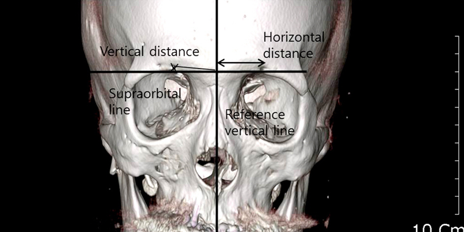
The study sample was composed of 87 adult patients with no history of trauma or ocular disease. The horizontal position of the supraorbital foramen or notch was recorded in relation to a vertical line defined by a reproducible hypothetical point, such as the nasion and mid-maxilla and the midpoint of the horizontal supraorbital plane. The distance and angle for each supraorbital foramen and notch were calculated from the defined vertical line. Furthermore, vertical distance from supraorbital plane, which was established using the highest points of both supraorbital rims, was obtained from the supraorbital foramen.
RESULTS
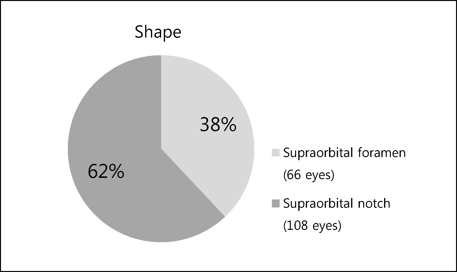
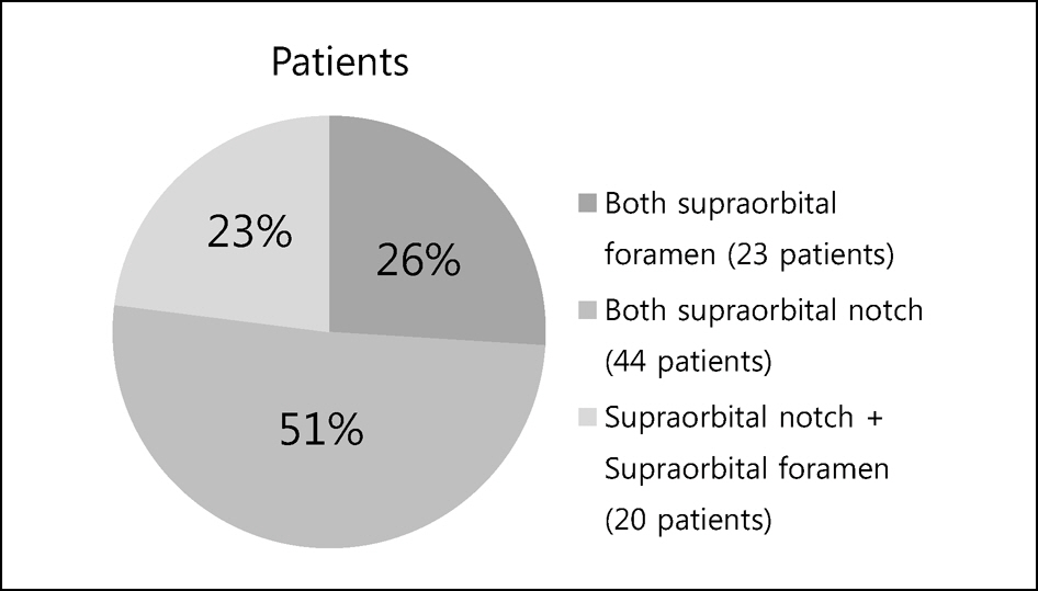
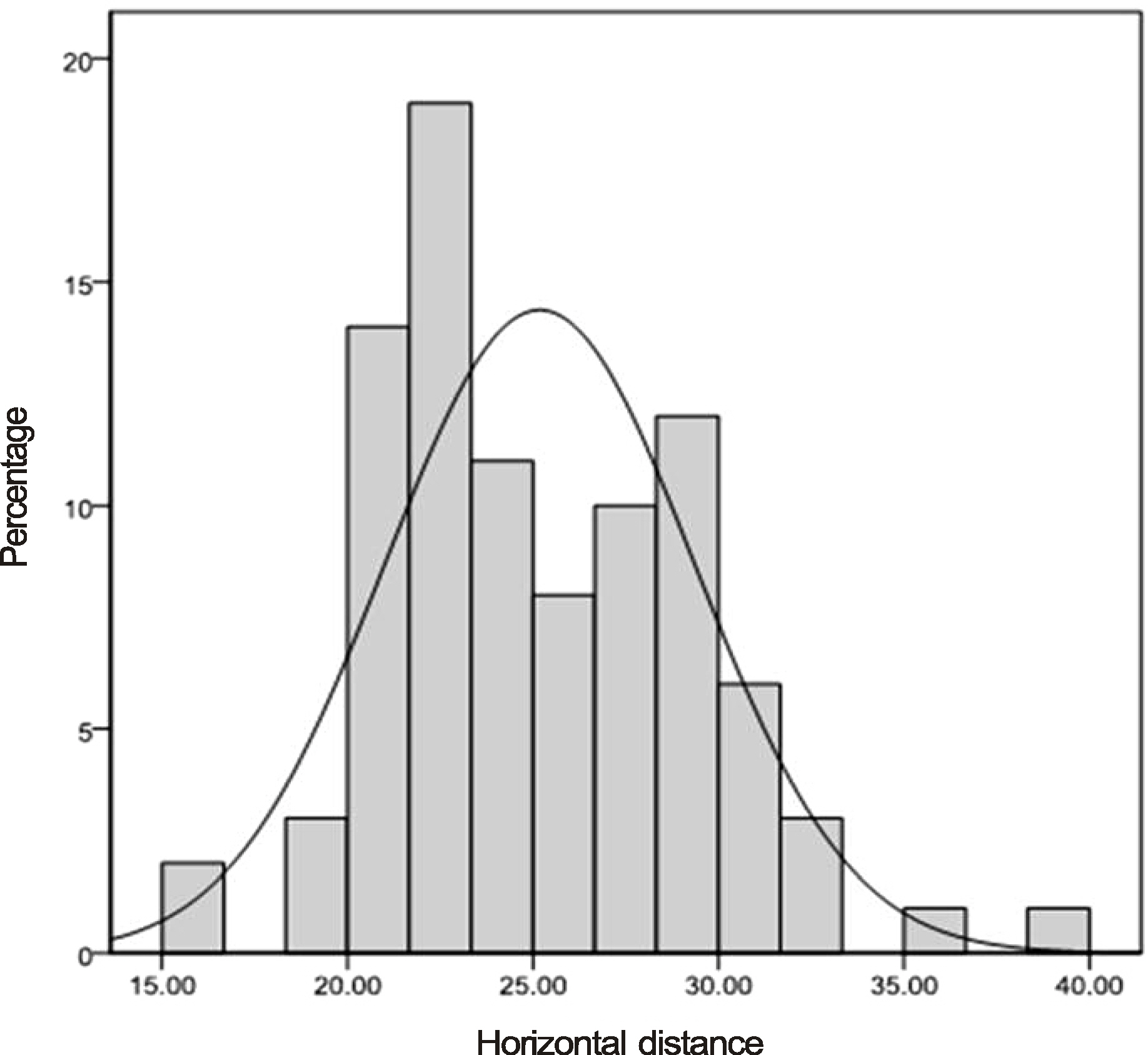
The mean age of the 87 patients was 45.44 +/- 8.34 years (range, 30-59 years). There were 66 eyes in the supraorbital notch and 108 eyes in the supraorbital foramen. There were no distributional differences between the 2 sides. The mean horizontal distance of both types was 23.95 +/- 3.93 mm (range, 16.41-38.94 mm). The horizontal distance of male patients was longer than the female patients (25.18 +/- 4.16 mm vs. 22.63 +/- 3.19 mm, p < 0.001, based on independent t-test) and the horizontal distance of supraorbital notch was shorter than the supraorbital foramen (22.59 +/- 3.18 mm vs. 26.18 +/- 4.04 mm, respectively, p < 0.001, based on independent t-test). The mean vertical distance and mean angles of the supraorbital foramen were 3.02 +/- 1.119 mm and 6.81 +/- 2.31 degrees (degrees), respectively.
CONCLUSIONS
The present study described the anatomical location of each supraorbital opening type in Korean adults. According to horizontal distance, a surgeon can avoid iatrogenic injury of the supraorbital neurovascular complex, especially during brow surgery. In addition, the anatomy can aid in targeting supraorbital neurovascular complex in cases of nerve block.
MeSH Terms
Figure
Reference
-
References
1. Booth AJ, Murray A, Tyers AG. The direct brow lift: efficacy, complications, and patient satisfaction. Br J Ophthalmol. 2004; 88:688–91.
Article2. Trivedi DJ, Shrimankar PS, Kariya VB, Pensi CA. A study of supraorbital notches and foramina in gujarati human skulls. NJIRM. 2010; 1:1–6.3. Beer GM, Putz R, Mager K, et al. Variations of the frontal exit of the supraorbital nerve: an anatomic study. Plast Reconstr Surg. 1998; 102:334–41.
Article4. Barker L, Naveed H, Adds PJ, Uddin JM. Supraorbital notch and foramen: positional variation and relevance to direct brow lift. Ophthal Plast Reconstr Surg. 2013; 29:67–70.5. Turhan-Haktanir N, Ayçiçek A, Haktanir A, Demir Y. Variations of supraorbital foramina in living subjects evaluated with multi-detector computed tomography. Head Neck. 2008; 30:1211–5.
Article6. Agthong S, Huanmanop T, Chentanez V. Anatomical variations of the supraorbital, infraorbital, and mental foramina related to gender and side. J Oral Maxillofac Surg. 2005; 63:800–4.
Article7. Webster RC, Gaunt JM, Hamdan US, et al. Supraorbital and supra-trochlear notches and foramina: anatomical variations and surgical relevance. Laryngoscope. 1986; 96:311–5.8. Saylam C, Ozer MA, Ozek C, Gurler T. Anatomical variations of the frontal and supraorbital transcranial passages. J Craniofac Surg. 2003; 14:10–2.
Article9. McCord CD, Codner MA, Hester TR. Eyelid surgery: Principles and techniques. New York: Lippincott-Raven;1995. p. 166–95.10. Ueda K, Harii K, Yamada A. Long-term follow-up study of brow-lift for treatment of facial paralysis. Ann Plast Surg. 1994; 32:166–70.
Article11. Cavalcanti MG, Rocha SS, Vannier MW. Craniofacial measurements based on 3D-CT volume rendering: implications for clinical applications. Dentomaxillofac Radiol. 2004; 33:170–6.
Article12. Waitzman AA, Posnick JC, Armstrong DC, Pron GE. Craniofacial skeletal measurements based on computed tomography: Part I. Accuracy and reproducibility. Cleft Palate Craniofac J. 1992; 29:112–7.
Article
- Full Text Links
- Actions
-
Cited
- CITED
-
- Close
- Share
- Similar articles
-
- Anatomic Characteristics of Supraorbital Foramina in Korean Using Three-Dimensional Model
- Anthropometric Analysis of Facial Foramina in Korean Population: A Three-Dimensional Computed Tomographic Study
- Confirmation of Supraorbital Nerve and Its Branch in the Supraorbital Notch with Ultrasound Guidance
- Anatomical structure of lingual foramen in cone beam computed tomography
- ANATOMICAL ASSESSMENT OF ACCESSORY MENTAL FORAMEN USING 3D CONE BEAM COMPUTED TOMOGRAPHY IN KOREAN


