Korean J Pain.
2013 Apr;26(2):130-134. 10.3344/kjp.2013.26.2.130.
Anatomic Characteristics of Supraorbital Foramina in Korean Using Three-Dimensional Model
- Affiliations
-
- 1Department of Anesthesiology and Pain Medicine, Seoul National University Bundang Hospital, Seongnam, Korea. ejchoi@snubh.org
- 2Department of Applied Mathematics and Statistics, State University of New York at Stony Brook, USA.
- KMID: 1978656
- DOI: http://doi.org/10.3344/kjp.2013.26.2.130
Abstract
- BACKGROUND
The aims of this study were to analyze the anatomic variations of supraorbital foramina/notches in Koreans and to compare the results with those of previous studies examining other races. We evaluated the three-dimensional computed tomography (3D-CT) images of human faces using multidetector computed tomography (MDCT).
METHODS
A total of 395 adults (232 men and 163 women) were enrolled and the 3D-CT images of their faces were reviewed in this study. In this study, the data from the images included the presence, shape, width and distance from the nasion to the supraorbital foramina/notches. ANOVA was used to assess the main effects of gender and side (right or left foramen/notch), and comparisons of the means were done by paired t-test.
RESULTS
The most common shapes in Koreans were a single notch (39.5%) on the right hand side and a single foramen (42.3%) on the left hand side. The incidence of a single foramen in Koreans was high compared to other races. The mean foramen diameter was 2.34 +/- 0.78 mm, and the mean distance from the nasion was 27.19 +/- 4.03 mm. The mean notch diameter was 3.37 +/- 1.71 mm, and the mean distance from the nasion was 23.42 +/- 2.45 mm.
CONCLUSIONS
This is the first study on the variations of supraorbital foramina/notches in Koreans using 3D-CT images of faces. The anatomic characteristics of the supraorbital foramina/notch will help in performing nerve blocks and maxillofacial surgery.
MeSH Terms
Figure
-
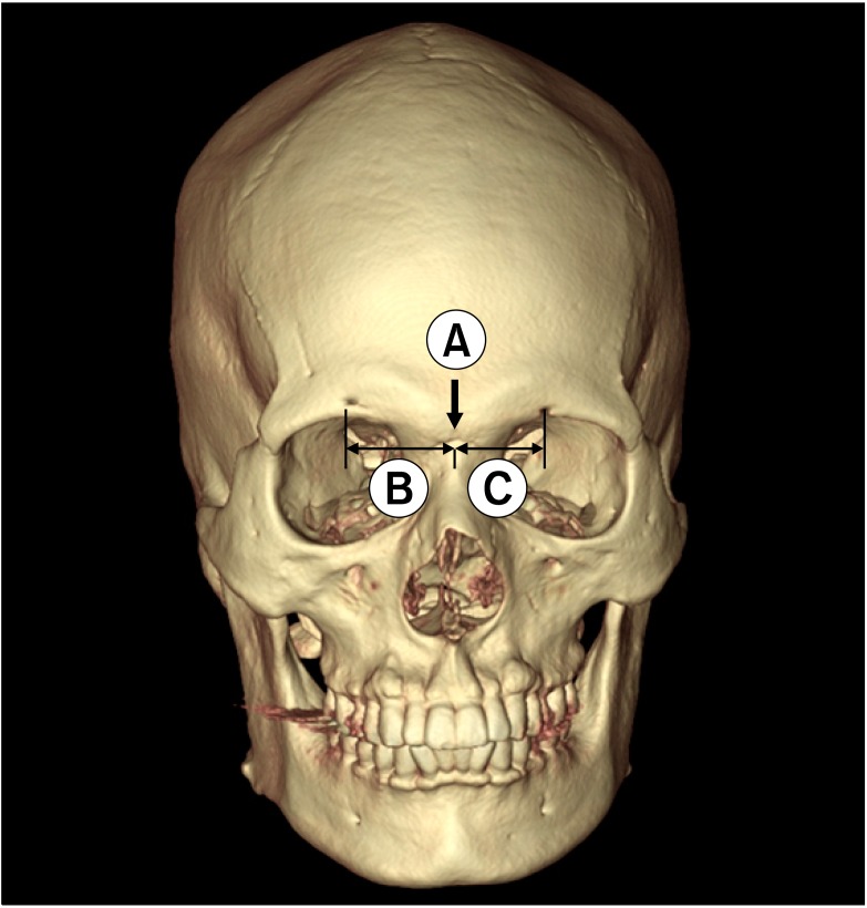
Fig. 1 Measurement methods for the three-dimensional computed tomography images. (A) Black arrow indicates nasion. (B) Distance from the center of the supraorbital foramen to nasion. (C) Distance from the center of the supraorbital notch to the nasion.

Fig. 2 Examples of anatomical variations. (A) Single notch (black arrow) on the right hand side and single foramen (open black arrow) on the left hand side. (B) None on both sides. (C) Double foramen (black arrows) on the left side.
Reference
-
1. Cutright B, Quillopa N, Schubert W. An anthropometric analysis of the key foramina for maxillofacial surgery. J Oral Maxillofac Surg. 2003; 61:354–357. PMID: 12618976.
Article2. Beer GM, Putz R, Mager K, Schumacher M, Keil W. Variations of the frontal exit of the supraorbital nerve: an anatomic study. Plast Reconstr Surg. 1998; 102:334–341. PMID: 9703067.
Article3. Caputi CA, Firetto V. Therapeutic blockade of greater occipital and supraorbital nerves in migraine patients. Headache. 1997; 37:174–179. PMID: 9100402.
Article4. Sjaastad O, Stolt-Nielsen A, Pareja JA, Fredriksen TA, Vincent M. Supraorbital neuralgia. On the clinical manifestations and a possible therapeutic approach. Headache. 1999; 39:204–212. PMID: 15613215.
Article5. Webster RC, Gaunt JM, Hamdan US, Fuleihan NS, Giandello PR, Smith RC. Supraorbital and supratrochlear notches and foramina: anatomical variations and surgical relevance. Laryngoscope. 1986; 96:311–315. PMID: 3951310.6. Agthong S, Huanmanop T, Chentanez V. Anatomical variations of the supraorbital, infraorbital, and mental foramina related to gender and side. J Oral Maxillofac Surg. 2005; 63:800–804. PMID: 15944977.
Article7. Chung MS, Kim HJ, Kang HS, Chung IH. Locational relationship of the supraorbital notch or foramen and infraorbital and mental foramina in Koreans. Acta Anat (Basel). 1995; 154:162–166. PMID: 8722516.
Article8. Cavalcanti MG, Haller JW, Vannier MW. Three-dimensional computed tomography landmark measurement in craniofacial surgical planning: experimental validation in vitro. J Oral Maxillofac Surg. 1999; 57:690–694. PMID: 10368094.
Article9. Turhan-Haktanir N, Ayçiçek A, Haktanir A, Demir Y. Variations of supraorbital foramina in living subjects evaluated with multidetector computed tomography. Head Neck. 2008; 30:1211–1215. PMID: 18642294.
Article10. Klein DS, Schmidt RE Jr. Chronic headache resulting from postoperative supraorbital neuralgia. Anesth Analg. 1991; 73:490–491. PMID: 1822965.
Article11. Evans RW, Pareja JA. Expert opinion. Supraorbital neuralgia. Headache. 2009; 49:278–281. PMID: 19222598.
- Full Text Links
-
- Actions
-
Cited
- CITED
-
- Close
- Share
-
- Similar articles
-
- Anthropometric Analysis of Facial Foramina in Korean Population: A Three-Dimensional Computed Tomographic Study
- Supraorbital nerve exits: positional variations and localization relative to surgical landmarks
- Anatomical Variant of Atlas : Arcuate Foramen, Occpitalization of Atlas, and Defect of Posterior Arch of Atlas
- Morphometric Analysis of the Supraorbital and Infraorbital Foramina Based on the Medial Canthus in Koreans
- Distribution of the lingual foramina in mandibular cortical bone in Koreans
