J Korean Ophthalmol Soc.
2015 Jan;56(1):13-18. 10.3341/jkos.2015.56.1.13.
Changes of the Meibomian Gland According to Age in the Normal Korean Population
- Affiliations
-
- 1Department of Ophthalmology, Kangnam Sacred Heart Hospital, Hallym University College of Medicine, Seoul, Korea. schinn@hanmail.net
- 2Department of Ophthalmology, Seoul National University Bundang Hospital, Seoul National University College of Medicine, Seongnam, Korea.
- 3Department of Ophthalmology, Seoul National University College of Medicine, Seoul, Korea.
- KMID: 2216148
- DOI: http://doi.org/10.3341/jkos.2015.56.1.13
Abstract
- PURPOSE
To examine the changes in meibomian glands associated with aging in a normal Korean population and to estimate the differences between the upper and lower eyelid in each age group.
METHODS
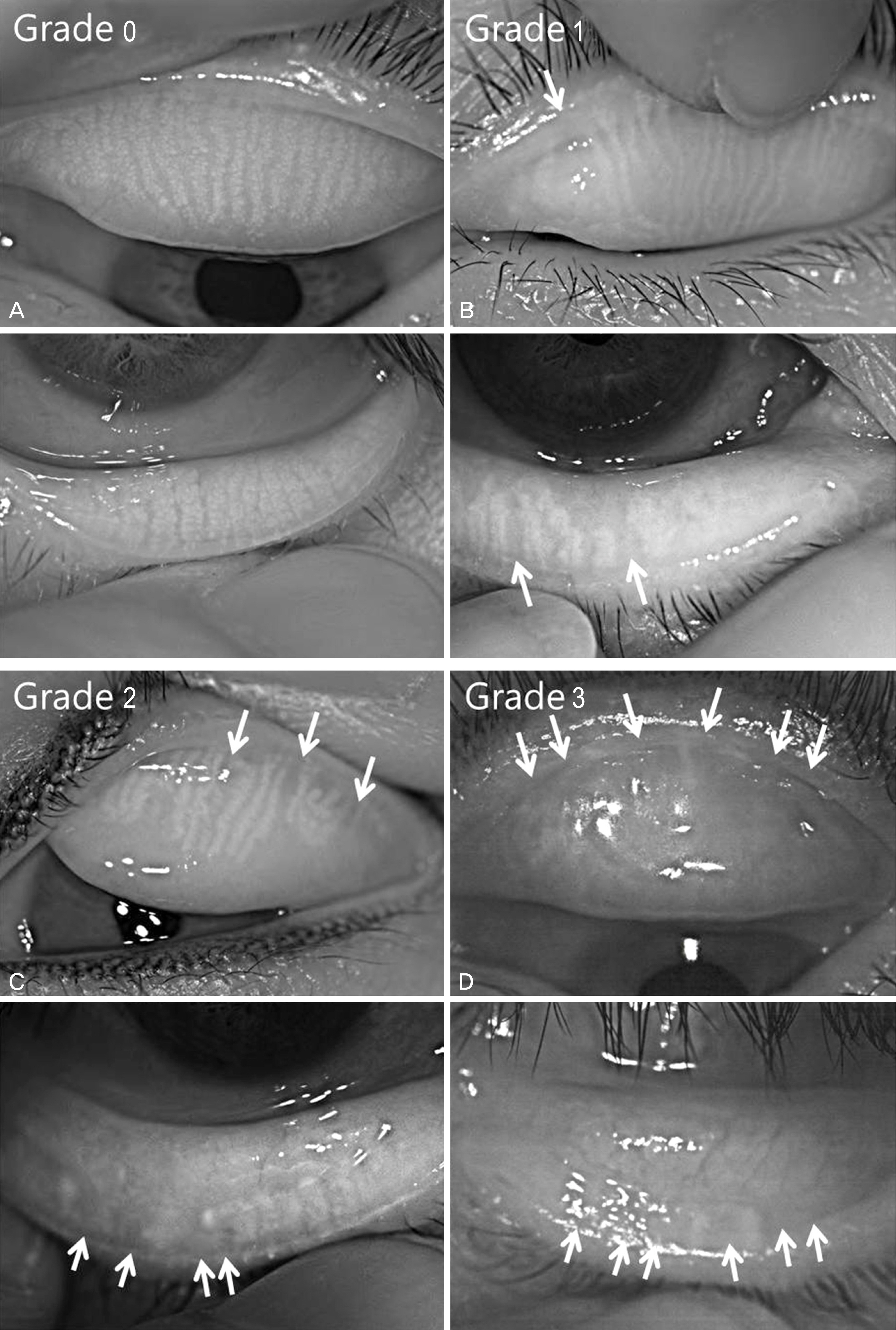
We performed meibography on adult subjects using an infrared charge-coupled device (CCD) camera. Each eyelid was scored based on the loss of meibomian glands, and the meiboscores of the upper and lower eyelids were summed to obtain a score for each eye. Meiboscores were evaluated according to age, sex, and upper and lower eyelid meiboscores in each age group.
RESULTS
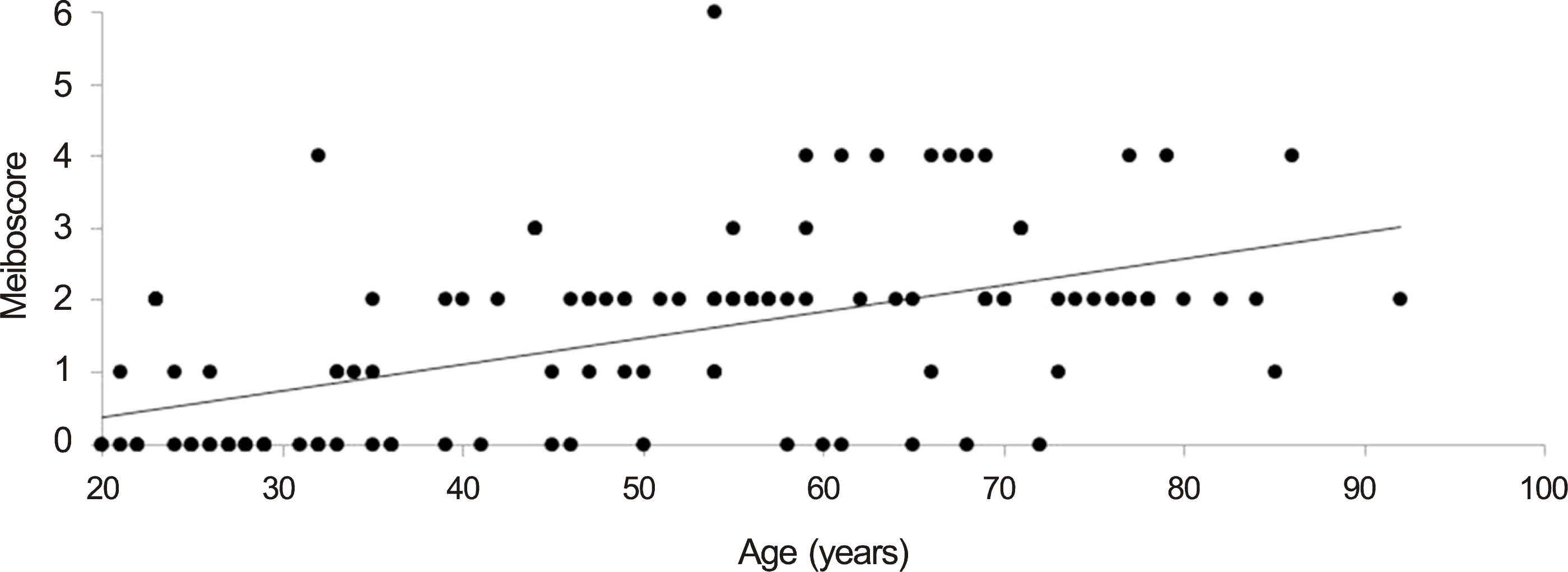
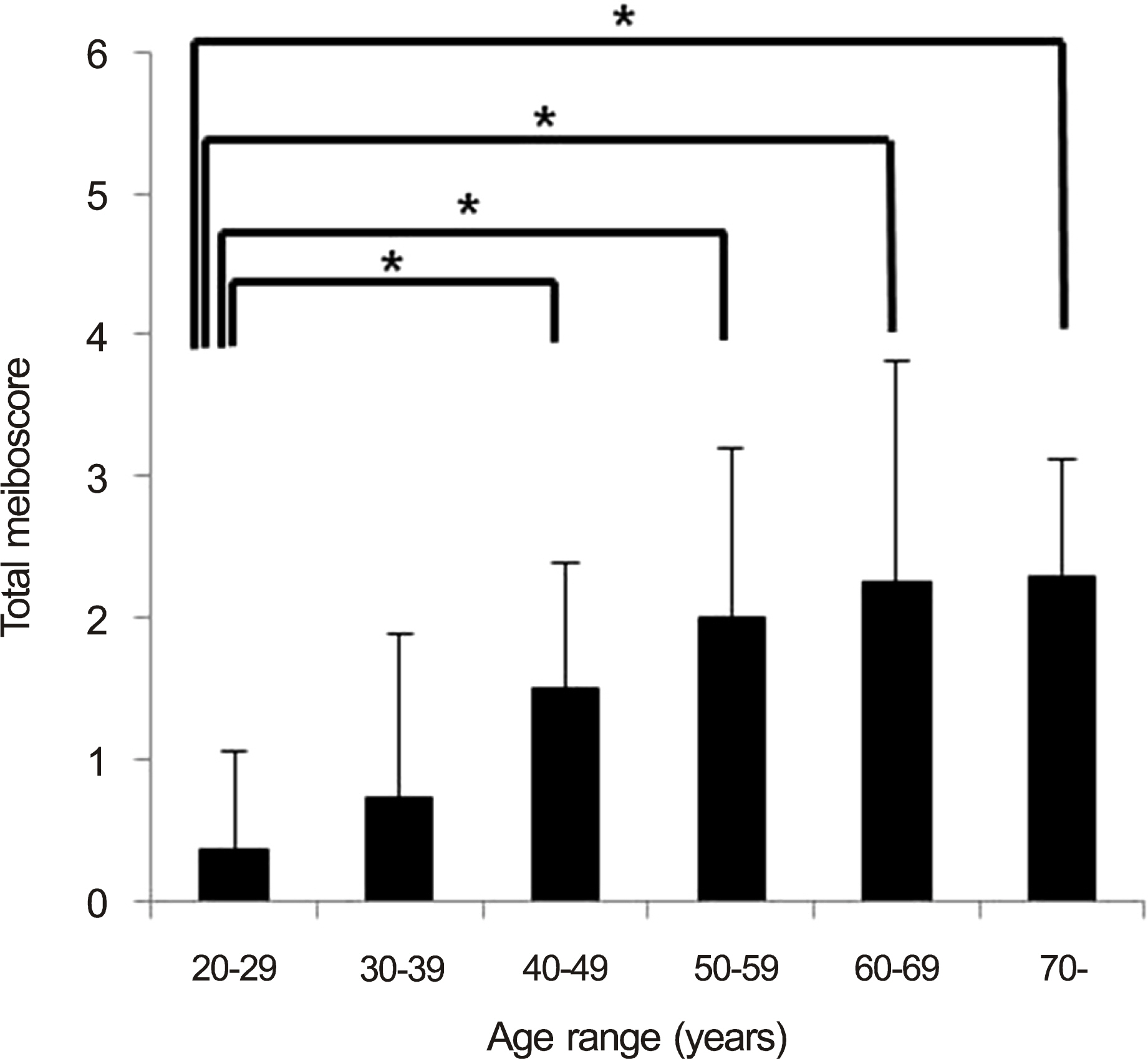
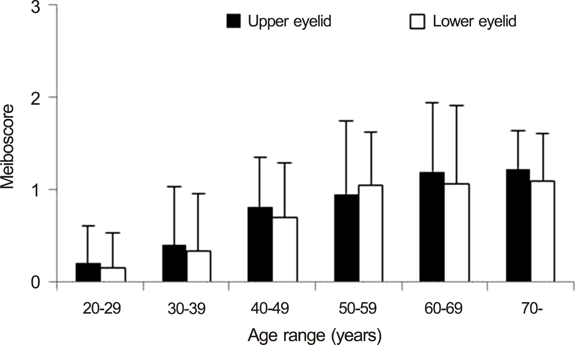
One hundred seventeen eyes of 117 people were enrolled in this study. The study subjects had an average age of 50.4 +/- 19.1 years (range, 20-92; male, 56; female, 61). There was a significant positive correlation between age and total meiboscore, upper and lower eyelid meiboscore (r = 0.578, p < 0.001; r = 0.550, p < 0.001; r = 0.524, p < 0.001). There were no significant differences in the meiboscores of the upper and lower eyelids in any age group, though meiboscores were significantly higher since 40 year-old group than 20 year-old group (p < 0.001, p < 0.001, p < 0.001, p < 0.001).
CONCLUSIONS
Based on noncontact meibography in normal adult eyes, the authors concluded that prevalence of changes in the meibomian glands was about 60% and changes in meibomian glands increase with age.
Keyword
Figure
Cited by 3 articles
-
Risk Factors for Structural Changes in Meibomian Gland in Thyroid Eye Disease
Minjae Kim, Sungwon Yang, Jinhwan Park, Hwa Lee, Sehyun Baek
J Korean Ophthalmol Soc. 2018;59(7):599-605. doi: 10.3341/jkos.2018.59.7.599.Meibography and Immunohistochemistric Structures in Animal Models
Hyun Kyu Lee, In Ho Woo, Boram Kang, Young Sub Eom, Su-Yeon Kang, Ho Sik Hwang, Hyo Myung Kim, Jong Suk Song
J Korean Ophthalmol Soc. 2018;59(1):9-16. doi: 10.3341/jkos.2018.59.1.9.Association between Dry Eye Questionnaires and Dry Eye Sign in Meibomian Gland Dysfunction
Jin A Kim, Si Hyung Lee
J Korean Ophthalmol Soc. 2020;61(2):138-145. doi: 10.3341/jkos.2020.61.2.138.
Reference
-
References
1. MISHIMA S, MAURICE DM. The oily layer of the tear film and evaporation from the corneal surface. Exp Eye Res. 1961; 1:39–45.
Article2. Nelson JD, Shimazaki J, Benitez-del-Castillo JM. . The international workshop on meibomian gland dysfunction: report of the definition and classification subcommittee. Invest Ophthalmol Vis Sci. 2011; 52:1930–7.
Article3. Foulks GN, Bron AJ. Meibomian gland dysfunction: a clinical scheme for description, diagnosis, classification, and grading. Ocul Surf. 2003; 1:107–26.
Article4. Mathers WD. Ocular evaporation in meibomian gland dysfunction and dry eye. Ophthalmology. 1993; 100:347–51.
Article5. Cho JH, Ahn Y. Assessment of meibomian gland dysfunction and comparison of the results of BUT and Schirmer test according to meibomian gland state. J Korean Ophthalmol Soc. 2000; 41:1875–82.6. Mathers WD, Daley T, Verdick R. Video imaging of the meibomian gland. Arch Ophthalmol. 1994; 112:448–9.
Article7. Nichols JJ, Berntsen DA, Mitchell GL, Nichols KK. An assessment of grading scales for meibography images. Cornea. 2005; 24:382–8.
Article8. Tomlinson A, Bron AJ, Korb DR. . The international workshop on meibomian gland dysfunction: report of the diagnosis subcommittee. Invest Ophthalmol Vis Sci. 2011; 52:2006–49.
Article9. Ibrahim OM, Matsumoto Y, Dogru M. . The efficacy, sensitivity, and specificity of in vivo laser confocal microscopy in the diagnosis of meibomian gland dysfunction. Ophthalmology. 2010; 117:665–72.
Article10. Hwang HS, Park CW, Joo CK. Novel noncontact meibography with anterior segment optical coherence tomography: Hosik meibography. Cornea. 2013; 32:40–3.11. Srinivasan S, Menzies K, Sorbara L, Jones L. Infrared imaging of meibomian gland structure using a novel keratograph. Optom Vis Sci. 2012; 89:788–94.
Article12. Arita R, Itoh K, Inoue K, Amano S. Noncontact infrared meibography to document age-related changes of the meibomian glands in a normal population. Ophthalmology. 2008; 115:911–5.
Article13. Tapie R. Biomicroscopic study of the glands of meibomius. Ann Ocul. 1977; 210:637–48.14. Yokoi N, Komuro A, Yamada H. . A newly developed vid-eo-meibography system featuring a newly designed probe. Jpn J Ophthalmol. 2007; 51:53–6.
Article15. Jester JV, Rife L, Nii D. . In vivo biomicroscopy and photography of meibomian glands in a rabbit model of meibomian gland dysfunction. Invest Ophthalmol Vis Sci. 1982; 22:660–7.16. Den S, Shimizu K, Ikeda T. . Association between meibomian gland changes and aging, sex, or tear function. Cornea. 2006; 25:651–5.
Article17. Norn M. Expressibility of meibomian secretion. Relation to age, lipid precorneal film, scales, foam, hair and pigmentation. Acta Ophthalmol (Copenh). 1987; 65:137–42.18. Pflugfelder SC, Tseng SC, Sanabria O. . Evaluation of subjective assessments and objective diagnostic tests for diagnosing tearfilm disorders known to cause ocular irritation. Cornea. 1998; 17:38–56.
Article19. Pult H, Riede-Pult B. Comparison of subjective grading and objective assessment in meibography. Cont Lens Anterior Eye. 2013; 36:22–7.
Article
- Full Text Links
- Actions
-
Cited
- CITED
-
- Close
- Share
- Similar articles
-
- A Case of Meibomian Gland Carcinoma
- Meibomian Gland Changes after Incision of Chalazions by Meibography
- Assessment of Meibomian Gland Dysfunction and Comparison of The Results of BUT and Schirmer Test According to Meibomian Gland State
- Two Cases Report Meibomian Gland Carcinoma
- Risk Factors of Meibomian Gland Loss in Dry Eye Disease


