Multiple Cerebral Infarctions with Neurological Symptoms and Ophthalmic Artery Occlusion after Filler Injection
- Affiliations
-
- 1Department of Ophthalmology, Kangdong Sacred Heart Hospital, Hallym University College of Medicine, Seoul, Korea. sungpyo@hanafos.com
- KMID: 2216015
- DOI: http://doi.org/10.3341/jkos.2015.56.2.285
Abstract
- PURPOSE
To report a case of visual loss, side weakness and facial palsy due to ophthalmic artery occlusion with diffuse multiple cerebral infarctions after injection of hyaluronic acid.
CASE SUMMARY
A 50-year-old female visited our clinic for visual loss in the left eye after filler injection in the glabella. Her best corrected visual acuity was 1.0 in the right eye and hand motion in the left eye. The intraocular pressure was 8 mm Hg in the right eye and 14 mm Hg in the left eye. In the left eye, there was abnormal pupillary light reflex and complete extra-ocular muscles palsy with blepharoptosis. A pale retina with a cherry-red-spot also appeared in the left fundus. A central retinal artery occlusion was observed on fluorescein angiography and brain magnetic resonance imaging showed multiple cerebral infarctions at the frontal, temporal, parietal and occipital lobes. Four days later, the motor weakness was aggravated and dysarthria and aphasia became worse. According to symptoms, a hemorrhagic transformation in subacute infarctions developed based on brain computed tomography. After 3 months of follow up, the visual acuity in the left eye was no light perception. However, the general conditions including ophthalmoplegia and motor weakness were improved.
Keyword
MeSH Terms
-
Aphasia
Blepharoptosis
Brain
Cerebral Infarction*
Dysarthria
Facial Paralysis
Female
Fluorescein Angiography
Follow-Up Studies
Hand
Humans
Hyaluronic Acid
Infarction
Intraocular Pressure
Magnetic Resonance Imaging
Middle Aged
Muscles
Occipital Lobe
Ophthalmic Artery*
Ophthalmoplegia
Paralysis
Rabeprazole
Reflex
Retina
Retinal Artery Occlusion
Visual Acuity
Hyaluronic Acid
Figure
-
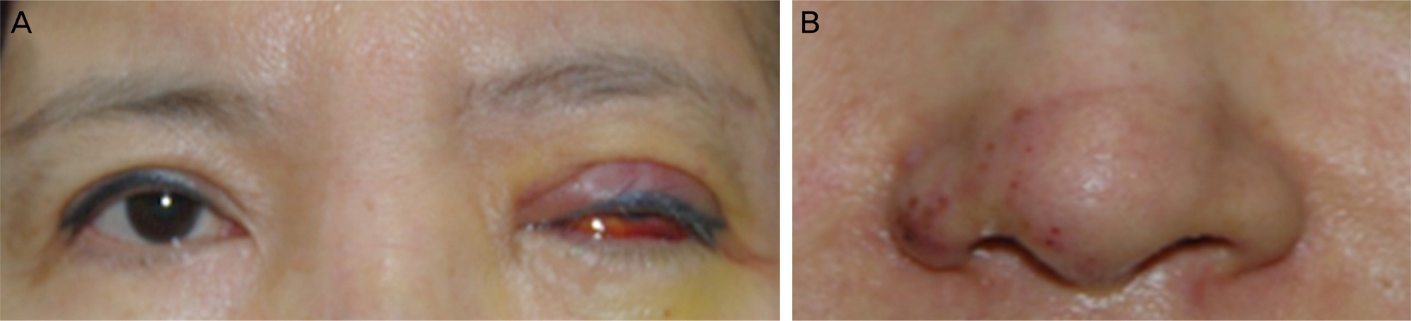
Figure 1. (A) Initial facial photographs show erythematous left eyelid swellings with blepharoptosis, severe conjunctival chemosis, subconjunctival hemorrhage in the left eye and (B) purpura like rash at nasal ala and forehead.
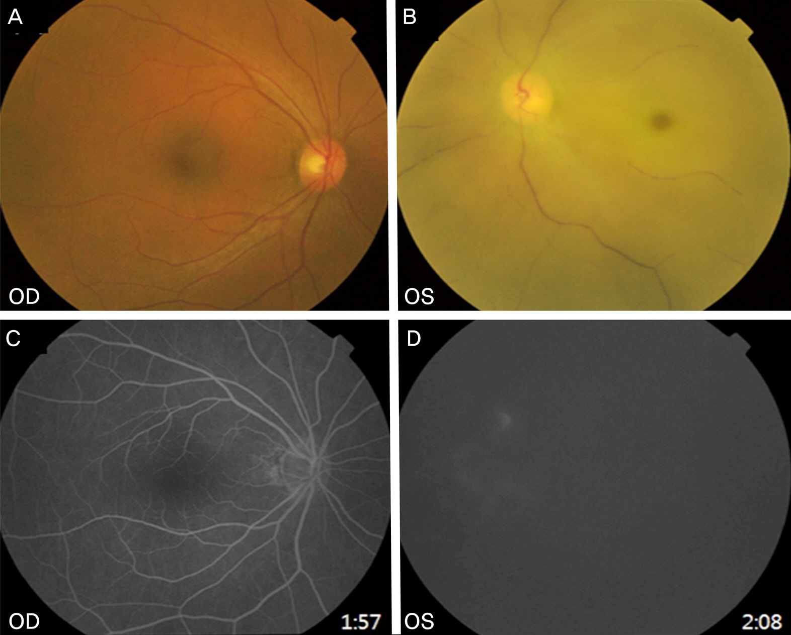
Figure 2. (A) Fundus pho- tographs show normal in right eye, (B) but pale retina and thin vessels with cherry red spot in the left eye. (C) Fundus fluorescein angiographs show normal in the right eye, (D) non- perfusion of retina and choroid in the left eye about 2 minute after dye injection.
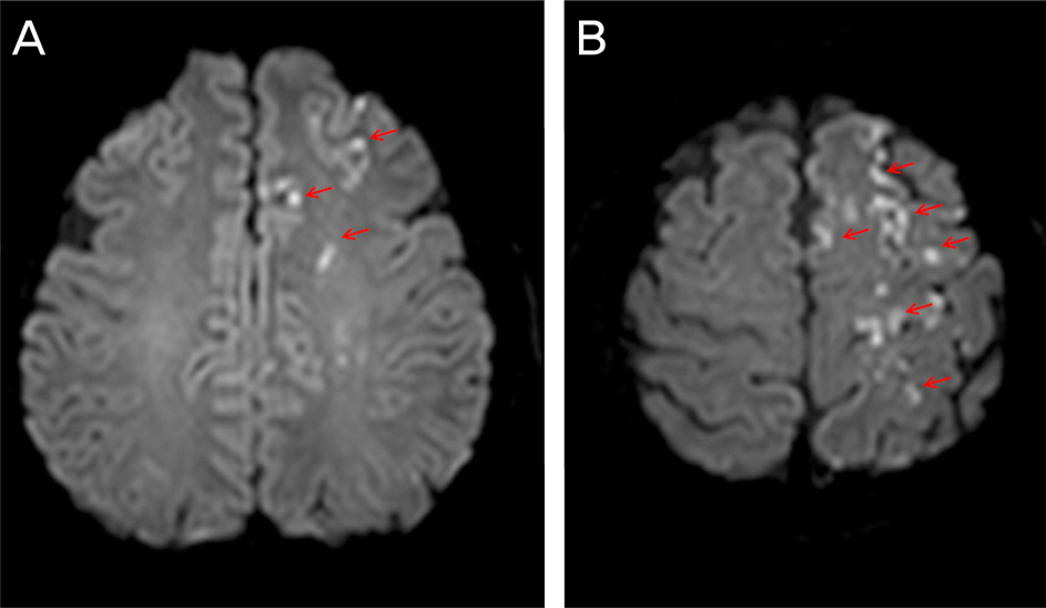
Figure 3. (A, B) Brain diffuse MRI (axial view) shows multiple high signal intensity lesions on left cerebral hemisphere (frontal, temporal, occipital, parietal lobes) which were suspected recent infarctions of middle cerebral artery territory (arrows).
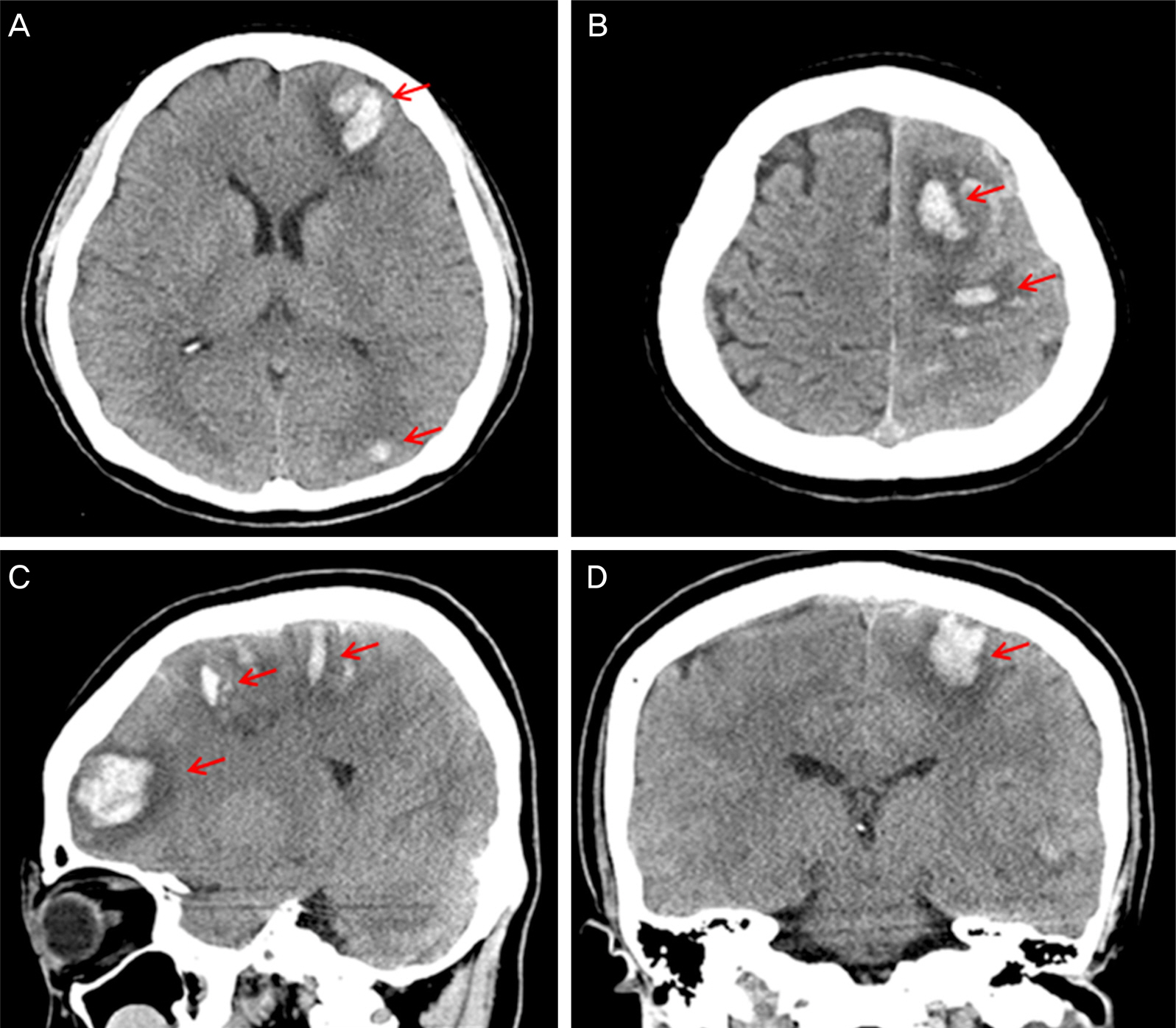
Figure 4. (A, B: axial view), (C: sagittal view), (D: coronal view) 4 days after filler injection, brain CT shows multiple hyper-dense hemorrhages (arrows) at left cerebral hemisphere (fro- ntal, temporal, occi-pital, parietal lobes) which mean hemorrhagic transfor- mation in subacute infarct- ions.
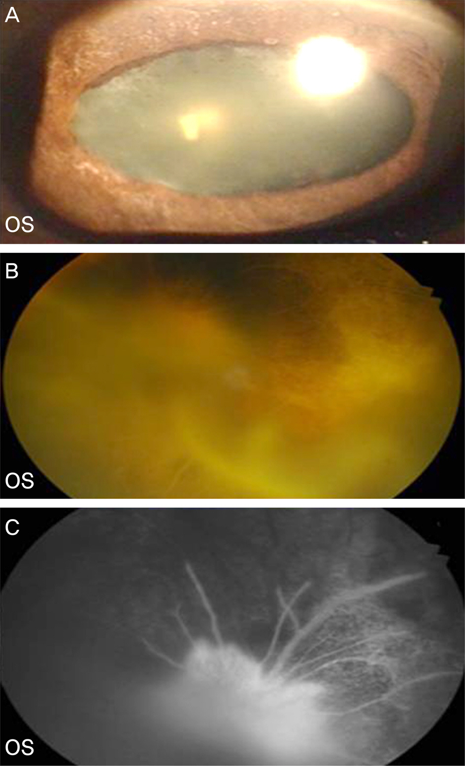
Figure 5. 3 months after filler injection (A) anterior segment photograph shows posterior synechiae and oval-shape fixed pupil. (B) Fundus photograph shows fibrovasular membrane around the disc and posterior pole. (C) Fundus fluorescein an-giograph shows hyperfluorescence because of fibrovascular membrane about 1 minute after dye injection.
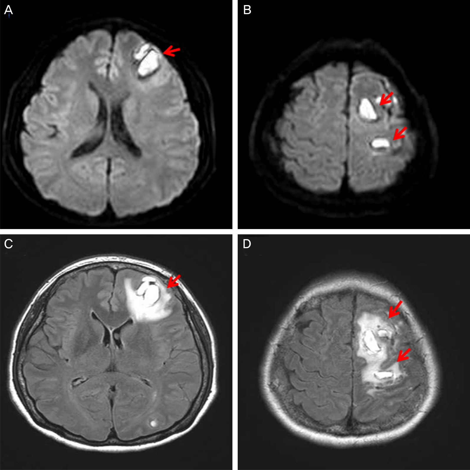
Figure 6. 3 months after filler injection, (A, B: difuseion MRI), (C, D: flare MRI) there’s no significant change compare to earlier image (Fig. 5) (arrows).
Cited by 1 articles
-
A Case of Orbital Apex Syndrome with Central Retinal Artery and Vein Occlusion Following Trauma
Mirinae Jang, Sang-Yoon Lee, Hye Jin Lee, Eun Kyoung Lee
J Korean Ophthalmol Soc. 2018;59(3):295-300. doi: 10.3341/jkos.2018.59.3.295.
Reference
-
References
1. Edwards PC, Fantasia JE. Review of long-term adverse effects as-sociated with the use of chemically-modified animal and non-animal source hyaluronic acid dermal fillers. Clin Interv Aging. 2007; 2:509–19.
Article2. Lee CM, Hong IH, Park SP. Ophthalmic artery obstruction and cerebral infarction following periocular injection of autologous fat. Korean J Ophthalmol. 2011; 25:358–61.
Article3. Park KH, Kim YK, Woo SJ. . Iatrogenic occlusion of the oph-thalmic artery after cosmetic facial filler injections: a national survey by the Korean Retina Society. JAMA Ophthalmol. 2014; 132:714–23.4. Sahin I, Isik S. Blindness following cosmetic injections of the face. Plast Reconstr Surg. 2012; 130:738e. author reply 738e-740e.
Article5. Paik DW, Jang IB, Kim JS. . A case of visual loss and oph-thalmoplegia following injection of hyaluronic acid into the glabella. J Korean Ophthalmol Soc. 2013; 54:971–6.
Article6. Eun YS, Cho SH, Lee JD, Kim HS. Periorbital lipogranuloma related to filler migration: a rare complication of facial fillers. J Cosmet Laser Ther. 2014; 16:149–50.
Article7. Banh K. Facial ischemia after hyaluronic acid injection. J Emerg Med. 2013; 44:169–70.
Article8. Tracy L, Ridgway J, Nelson JS. . Calcium hydroxylapatite associated soft tissue necrosis: a case report and treatment guideline. J Plast Reconstr Aesthet Surg. 2014; 67:564–8.
Article9. Park SW, Woo SJ, Park KH. . Iatrogenic retinal artery occlusion caused by cosmetic facial filler injections. Am J Ophthalmol. 2012; 154:653–62.e1.
Article10. Park SJ, Woo SJ, Park KH. . Partial recovery after intraarterial pharmacomechanical thrombolysis in ophthalmic artery occlusion following nasal autologous fat injection. J Vasc Interv Radiol. 2011; 22:251–4.
Article11. Sussman ES, Connolly ES Jr. Hemorrhagic transformation: a review of the rate of hemorrhage in the major clinical trials of acute ischemic stroke. Front Neurol. 2013; 4:69.
Article12. Khatri R, McKinney AM, Swenson B, Janardhan V. Blood-brain barrier, reperfusion injury, and hemorrhagic transformation in acute ischemic stroke. Neurology. 2012; 79(13 Suppl 1):S52–7.
Article13. Yassi N, Parsons MW, Christensen S. . Prediction of poststroke hemorrhagic transformation using computed tomography perfusion. Stroke. 2013; 44:3039–43.
Article
- Full Text Links
-
- Actions
-
Cited
- CITED
-
- Close
- Share
-
- Similar articles
-
- Hemorrhagic Stroke and Blindness after Hyaluronic Acid/Polylactic Acid Filler Injection
- Multifocal Retinal and Ciliary Artery Occlusion with Submacular Hemorrhage Following Cosmetic Facial Filler Injection
- Cerebral Angiographic Findings of Cosmetic Facial Filler-related Ophthalmic and Retinal Artery Occlusion
- Ophthalmic Artery Occlusion After Carotid Revascularization
- Central Retinal Artery Occlusion after Filler Injection for Upper Lid Retraction
