Comparative Analysis of Corneal Refraction and Astigmatism Measured with Autokeratometer, IOL Master, and Topography
- Affiliations
-
- 1Department of Ophthalmology, Seoul National University College of Medicine, Seoul, Korea. kmk9@snu.ac.kr
- 2Laboratory of Corneal Regenerative Medicine and Ocular Immunology, Seoul Artificial Eye Center, Seoul National University Hospital Clinical Research Institute, Seoul, Korea.
- 3Seoul National University Bundang Hospital, Seongnam, Korea.
- KMID: 2215145
- DOI: http://doi.org/10.3341/jkos.2011.52.12.1427
Abstract
- PURPOSE
To comparatively analyze the repeatability and consistency between different methods of measuring corneal refraction and astigmatism in order to precisely determine the toric intraocular lens target.
METHODS
The medical records of 254 eyes of 192 persons were retrospectively reviewed to compare the repeatability of corneal refraction measured with autokeratometer, IOL Master, and topography. The axis and the amount of the astigmatism were compared between these methods. The differences between the estimated spherical equivalents using measured corneal refractive values and the actual spherical equivalents after cataract surgery were compared to evaluate the expected astigmatic error of each measurement.
RESULTS
The average corneal refraction was greater with topography than with IOL Master or autokeratometer. Astigmatism calculated with IOL Master was greater than that measured with topography or autokeratometer. The mean coefficient of variation for mean corneal refraction was 0.19% with autokeratometer, which was smaller than that with IOL Master or topography. In patients with more than 1.5D of astigmatism, there were no significant differences in the axis measured by each instrument. The expected spherical error in IOL calculation was smaller with the measured values from IOL Master and autokeratometer than were those with topography.
CONCLUSIONS
The repeatability of measurements for corneal refraction and astigmatism was significantly higher using the autokeratometer and IOL Master, with the highest astigmatic value observed with the IOL master. The axis of astigmatism for each method was consistent in the patients with more than 1.5D of astigmatism.
Keyword
MeSH Terms
Figure
-
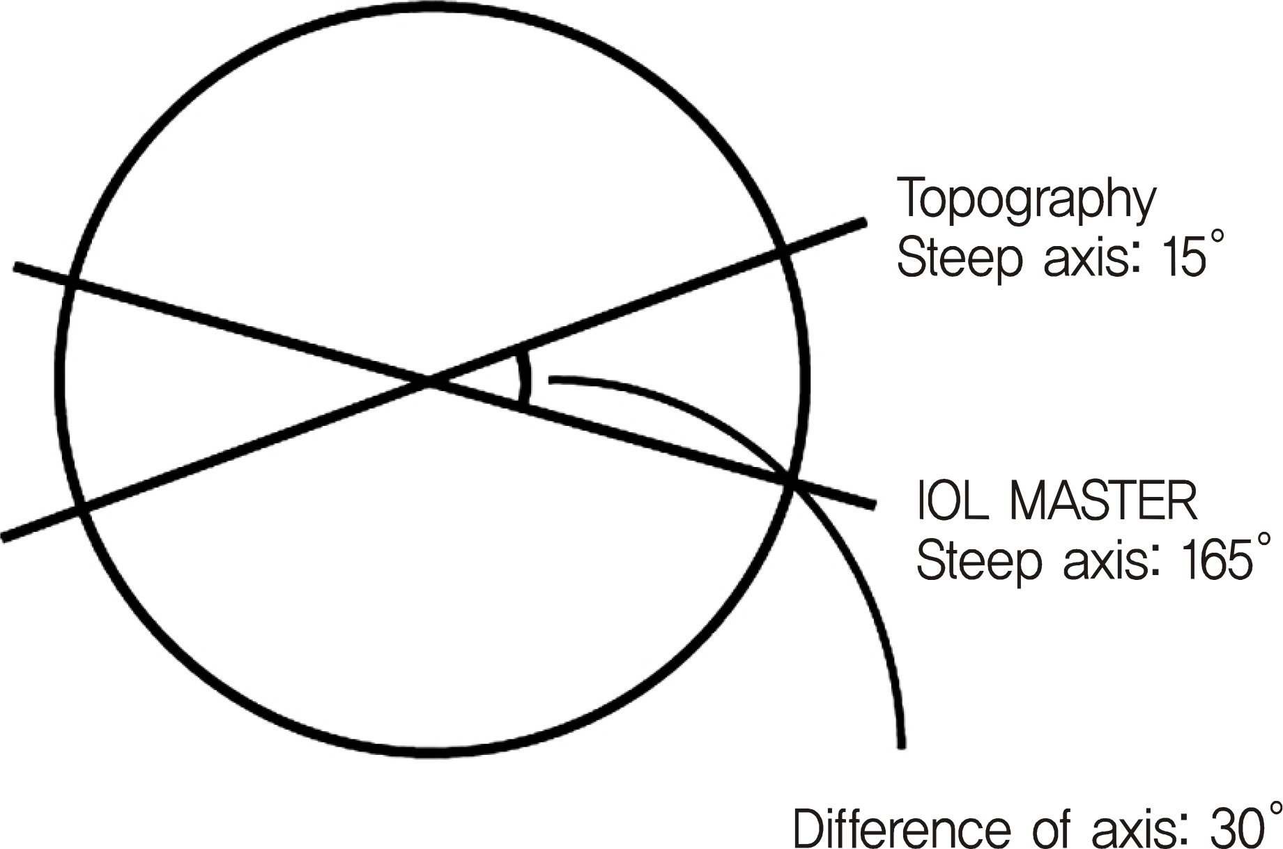
Figure 1. Difference of steep axis. For example, if the steep axis measured by topography is 15° and that measured by IOL Master is 165°, the difference of axis is 30°, neither 150° nor −30°.
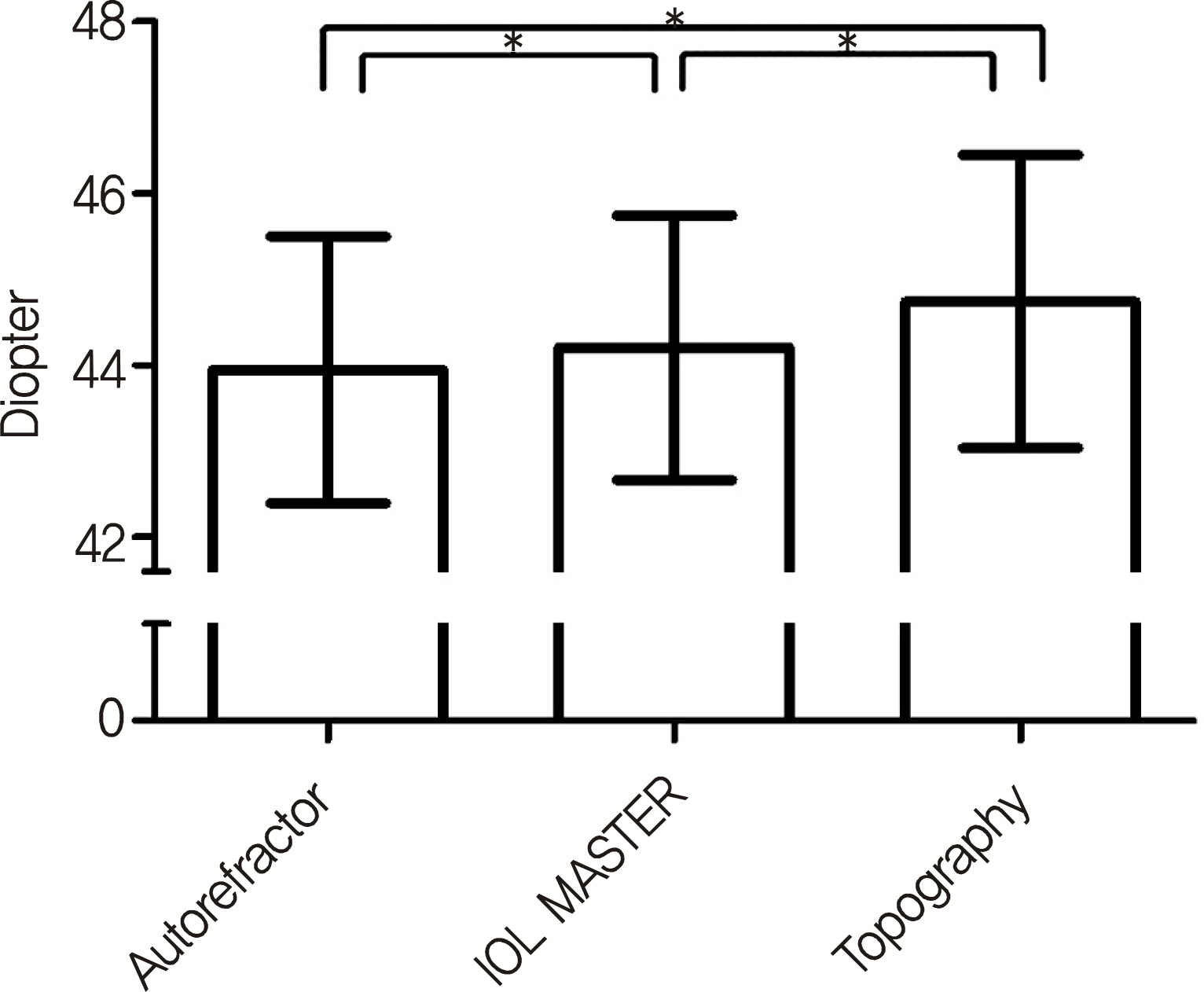
Figure 2. Difference between mean corneal refraction measured by autokeratometer, IOL Master, and topography. Note that these 3 values are significantly different (p < 0.001, paired t-test). ∗ p < 0.05.
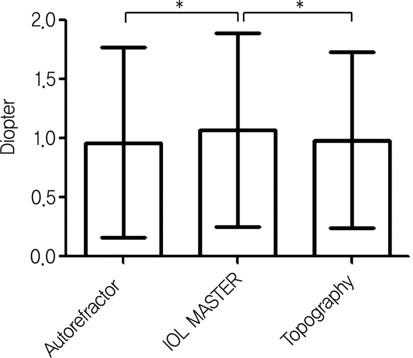
Figure 3. Difference between corneal astigmatism measured by autokeratometer, IOL Master, and topography. Note that astigmatism is measured more by IOL Master than autokeratometer and topography (respectively p = 0.003, p = 0.005, paired t-test). ∗ p < 0.05.
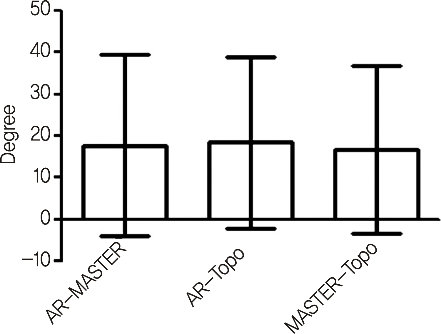
Figure 4. Difference between steep axis. Note that there are no statistically difference (p > 0.05, student's t-test).
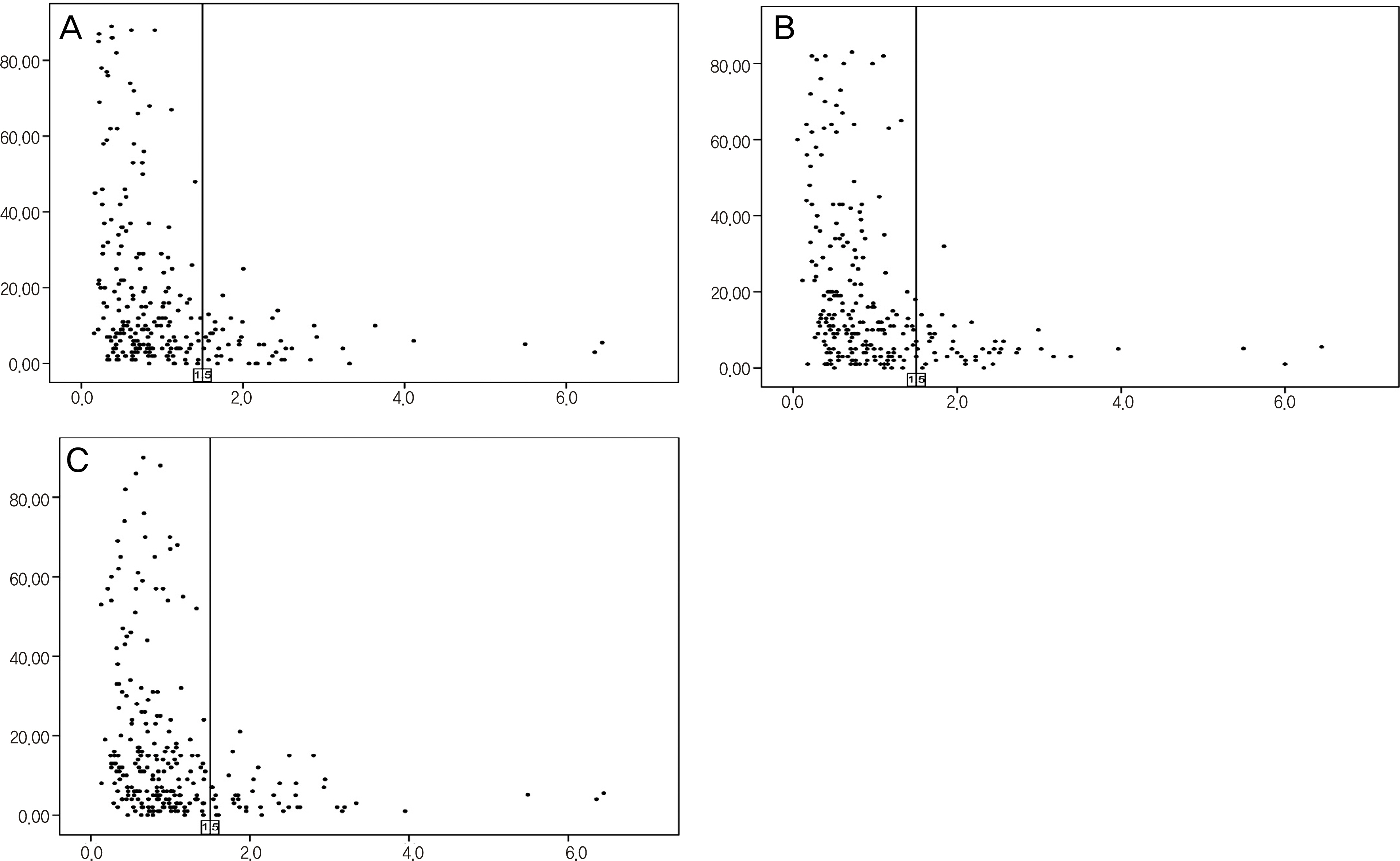
Figure 5. Difference of steep axis are scattered according to corneal astigmatism. Note that when the astigmatism is more than 1.5 diopters (vertical line), the difference is small and comparable.(A) Autokeratometer & IOL Master. (B) Autokeratometer & topography. (C) IOL Master & topography.
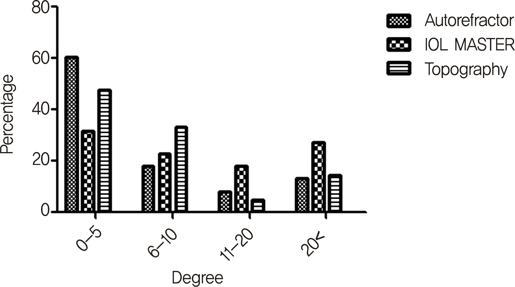
Figure 6. Difference of steep axis measured twice by the same method. Note that the group, greater than 11 degrees, are more common when measured by IOL Master (p = 0.033, chi-square).
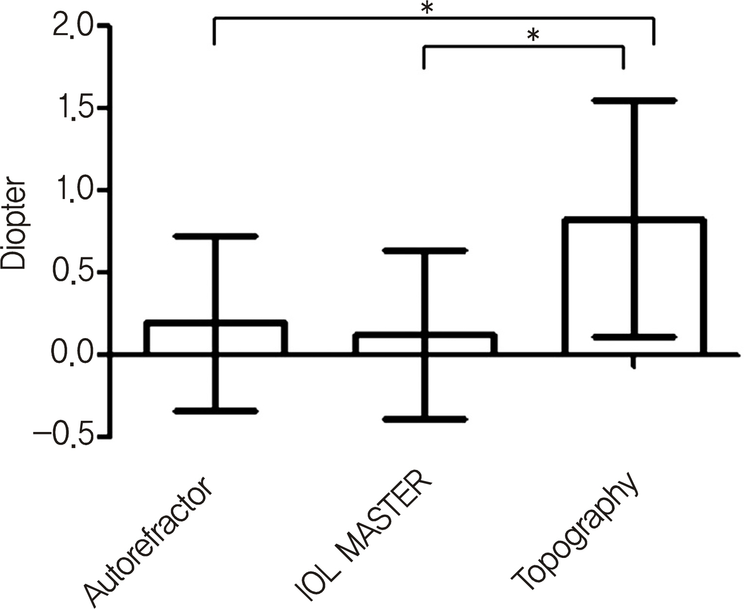
Figure 7. Comparison of expected spherical error (actual refraction minus predicted refraction) of IOL calculation measured by autokeratometer, IOL Master, and topography. IOL prediction is more accurate measured by autokeratometer and IOL Master, than by topography (Autorekeratometer vs. IOL Master p = 0.152, autokeratometer vs. topography p<0.001, IOL Master vs. topography p < 0.001, paired t-test). ∗ p < 0.05.
Cited by 5 articles
-
Comparative Analysis of Corneal Refractive Power Measured with AL-Scan®, Autokeratometer, and Pentacam®
Sung Jin Park, Sung Hyup Lim, Ho Young Lee
J Korean Ophthalmol Soc. 2014;55(7):984-990. doi: 10.3341/jkos.2014.55.7.984.Effect of Toric Intraocular Lens Implantation on Astigmatism in Cataract Surgery
Yong Jae Cha, Mee Kum Kim, Won Ryang Wee
J Korean Ophthalmol Soc. 2015;56(10):1544-1551. doi: 10.3341/jkos.2015.56.10.1544.Comparision of Corneal Refractive Power Measured with Opitcal Low-coherence Reflectometry, Autokeratometer, and Topography in Children
Jae Gyun Jeung, Gi Hyun Bae
J Korean Ophthalmol Soc. 2016;57(10):1535-1541. doi: 10.3341/jkos.2016.57.10.1535.Astigmatic Correlation between the Automated Refractometry and Dual Scheimpflug Analyzer in Pseudophakic Eyes
Seung Hun Park, In Seok Song, Min Cheol Seong, Hee Yoon Cho, Min Ho Kang
J Korean Ophthalmol Soc. 2016;57(3):361-368. doi: 10.3341/jkos.2016.57.3.361.Age-related Changes in Anterior, Posterior Corneal Astigmatism in a Korean Population
Yoon Seob Sim, Soon Won Yang, Yu Li Park, Kyung Sun Na, Hyun Seung Kim
J Korean Ophthalmol Soc. 2017;58(8):911-915. doi: 10.3341/jkos.2017.58.8.911.
Reference
-
References
1. Bellucci R. Multifocal intraocular lenses. Curr Opin Ophthalmol. 2005; 16:33–7.
Article2. Chang DF. Early rotational stability of the longer Staar toric intraocular lens: fifty consecutive cases. J Cataract Refract Surg. 2003; 29:935–40.3. Sanders DR, Gills JP, Martin RG. When keratometric measurements do not accurately reflect corneal topography. J Cataract Refract Surg. 1993; 19(Suppl):131–5.
Article4. Gormley DJ, Gersten M, Koplin RS, Lubkin V. Corneal modeling. Cornea. 1988; 7:30–5.
Article5. Potvin R, Fonn D, Sorbara L. In vivo comparison of corneal topography and keratometry systems. Int Contact Lens Clin. 1996; 23:20–6.
Article6. Shirayama M, Wang L, Koch DD, Weikert MP. Comparison of ac-curacy of intraocular lens calculations using automated keratometry, a Placido-based corneal topographer, and a combined Placido-based and dual Scheimpflug corneal topographer. Cornea. 2010; 29:1136–8.7. Butcher JM, O'Brien C. The reproducibility of biometry and keratometry measurements. Eye (Lond). 1991; 5:708–11.
Article8. Karabatsas CH, Cook SD, Papaefthymiou J, et al. Clinical evaluation of keratometry and computerised videokeratography: intraobserver and interobserver variability on normal and astigmatic corneas. Br J Ophthalmol. 1998; 82:637–42.
Article9. Hwang JS, Lee JH. Comparison of the IOL Master® and A-scan ultrasound: Refractive results of 96 consecutive cases. J Korean Ophthalmol Soc. 2007; 48:27–32.10. McKenzie RM, Kim YY, Bishop RW, Gaffney RJ. The collection and analysis of 2,3-dimethyl-2,3-dinitrobutane vapor. AIHA J. 1998; 59:388–92.
Article11. Forkman FJ. Coefficients of Variation – An Approximate F-Test. [Licentiate Thesis]. Uppsala: Swedish University of Agricultural Sciences;2005. 3.12. Vogel A, Dick HB, Krummenauer F. Reproducibility of optical biometry using partial coherence interferometry: intraobserver and interobserver reliability. J Cataract Refract Surg. 2001; 27:1961–8.
Article13. Shirayama M, Wang L, Weikert MP, Koch DD. Comparison of corneal powers obtained from 4 different devices. Am J Ophthalmol. 2009; 148:528–35.e1.
Article14. Kawamorita T, Uozato H, Kamiya K, et al. Repeatability, reproducibility, and agreement characteristics of rotating Scheimpflug photography and scanning-slit corneal topography for corneal power measurement. J Cataract Refract Surg. 2009; 35:127–33.
Article15. Nemeth J, Fekete O, Pesztenlehrer N. Optical and ultrasound measurement of axial length and anterior chamber depth for intraocular lens power calculation. J Cataract Refract Surg. 2003; 29:85–8.16. Huynh SC, Mai TQ, Kifley A, et al. An evaluation of keratometry in 6-year-old children. Cornea. 2006; 25:383–7.
Article17. Elbaz U, Barkana Y, Gerber Y, et al. Comparison of different techniques of anterior chamber depth and keratometric measurements. Am J Ophthalmol. 2007; 143:48–53.
Article
- Full Text Links
-
- Actions
-
Cited
- CITED
-
- Close
- Share
-
- Similar articles
-
- Clinical Reliability of IOL Master 700 in Measurement of Pupil Diameter and Corneal Curvature
- Pseudophakic Residual Astigmatism
- An Analysis of Correlation with Visual Acuity, Refractive Error and Corneal Astigmatism after Wearing of Reverse Geometry Lenses
- The Effect of Trabeculeetomy on Corneal Topography
- The Short-term Efficacy on Astigmatism Reduction According to Lenght and Site of Linear Clear Corneal Incision in Cataract Surgery
