Use of Fundus Autofluorescence Images to Evaluate the Progression of Geographic Atrophy: Two-Year Follow-Up Study
- Affiliations
-
- 1Department of Ophthalmology, Hallym University Kangdong Sacred Heart Hospital, Hallym University College of Medicine, Seoul, Korea. Sungpyo@hanafos.com
- KMID: 2215070
- DOI: http://doi.org/10.3341/jkos.2015.56.8.1195
Abstract
- PURPOSE
We evaluated the progression of geographic atrophy (GA) based on fundus autofluorescence (FAF) pattern and atrophy size using the fundus camera in non-exudative age-related macular degeneration (ARMD).
METHODS
We acquired FAF images in non-exudative ARMD patients over a 2-year period. According to The Fundus Autofluorescence in Age-related Macular Degeneration (FAM) study, FAF patterns of geographic atrophy were classified into 5 categories. Examiners quantified the areas of GA in FAF images and analyzed the progression of atrophy based on FAF pattern and atrophy size.
RESULTS
In 86 non-exudative ARMD eyes, elderly patients had faster progression rate of GA. The growth rates of GA were 1.51 mm2/year in 'Diffuse', 1.49 mm2/year in 'Banded', 1.05 mm2/year in 'Patchy', 0.59 mm2/year in 'Focal' and 0.16 mm2/year in 'None' pattern groups. In addition, the growth rate was 0.38 mm2/year in which initial the GA area was smaller than 1 disc area. This was the slowest progression rate among all categories according to initial GA area.
CONCLUSIONS
As a result of evaluating the progression of geographic atrophy using FAF over a 2-year period, the growth rate of GA was the fastest in the 'Diffuse' pattern group. Additionally, as the initial GA area became smaller, the progression of GA atrophy was slower (p < 0.002). Although limitations such as short follow-up period and measurement error of GA atrophy area using fundus photography were compensated, the results in the present study were similar to the outcomes of studies on progression of GA based on FAF pattern using the scanning laser ophthalmoscope over several years and the fundus camera for 1 year. In conclusion, the fundus camera is a useful tool for the prediction of long-term progression of GA in patients with non-exudative ARMD.
Keyword
MeSH Terms
Figure
-

Figure 1. Demarcation of geographic atrophy and calculation of its size. The examiners used a computer mouse to trace all areas of GA around the macula manually. Geographic atrophy was recognized as well-demarcated black areas corresponding to the dead or absent RPE. The computer software automates conversion of pixels to square mm based on the magnification factors of the imaging device. (A-1) None. (A-2) The atrophic area highlighted in red in the upper image. (B-1) Focal. (B-2) The atrophic area highlighted in red in the upper image. (C-1) Patchy. (C-2) The atrophic area highlighted in red in the upper image. (D-1) Banded. (D-2) The atrophic area highlighted in red in the upper image. (E-1) Diffuse. (E-2) The atrophic area highlighted in red in the upper image. GA = geographic atrophy; RPE = retinal pigment epithelium.
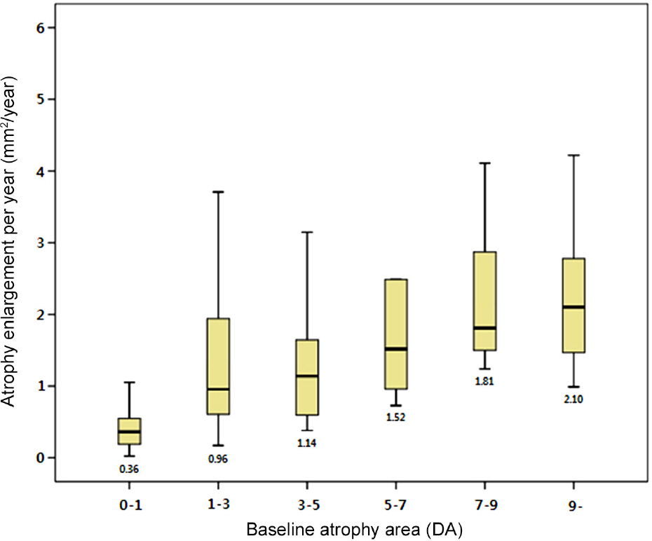
Figure 2. Eyes were classified depending on the total size of atrophy at baseline visit. Median atrophy enlargement per year is given below each box plot. Only the difference between the <1 DA group and the other groups was statistically significant ( p < 0.002). DA = disc area (2.54 mm2).
Reference
-
References
1. Bindewald-Wittich A, Han M, Schmitz-Valckenberg S. . Two-photon-excited fluorescence imaging of human RPE cells with a femtosecond Ti: Sapphire laser. Invest Ophthalmol Vis Sci. 2006; 47:4553–7.2. Bindewald A, Schmitz-Valckenberg S, Jorzik JJ. . Classification of abnormal fundus autofluorescence patterns in the junctional zone of geographic atrophy in patients with age related macular degeneration. Br J Ophthalmol. 2005; 89:874–8.
Article3. Delori FC, Dorey CK, Staurenghi G. . In vivo fluorescence of the ocular fundus exhibits retinal pigment epithelium lipofuscin characteristics. Invest Ophthalmol Vis Sci. 1995; 36:718–29.4. Jeong YJ, Hong IH, Chung JK. . Predictors for the progression of geographic atrophy in patients with age-related macular degen-eration: fundus autofluorescence study with modified fundus camera. Eye (Lond). 2014; 28:209–18.
Article5. Park KH, Song SJ, Lee WK. . The results of nation-wide regis-try of age-related macular degeneration in Korea. J Korean Ophthalmol Soc. 2010; 51:516–23.6. Topouzis F, Anastasopoulos E, Augood C. . Association of dia-betes with age-related macular degeneration in the EUREYE study. Br J Ophthalmol. 2009; 93:1037–41.
Article7. Youm DJ, Oh HS, Yu HG, Song SJ. The prevalence of vitreoretinal diseases in a screened Korean population 50 years and older. J Korean Ophthalmol Soc. 2009; 50:1645–51.
Article8. Friedman DS, O'Colmain BJ, Munoz B. . Prevalence of age-re-lated macular degeneration in the United States. Arch Ophthalmol. 2004; 122:564–72.
Article9. Holz FG, Bellmann C, Margaritidis M. . Patterns of increased in vivo fundus autofluorescence in the junctional zone of geo-graphic atrophy of the retinal pigment epithelium associated with age-related macular degeneration. Graefes Arch Clin Exp Ophthalmol. 1999; 237:145–52.
Article10. Sunness JS. The natural history of geographic atrophy, the ad-vanced atrophic form of age-related macular degeneration. Mol Vis. 1999; 5:25.11. Holz FG, Bindewald-Wittich A, Fleckenstein M. . Progression of geographic atrophy and impact of fundus autofluorescence pat-terns in age-related macular degeneration. Am J Ophthalmol. 2007; 143:463–72.
Article12. Holz FG, Schutt F, Kopitz J. . Inhibition of lysosomal degrada-tive functions in RPE cells by a retinoid component of lipofuscin. Invest Ophthalmol Vis Sci. 1999; 40:737–43.
- Full Text Links
-
- Actions
-
Cited
- CITED
-
- Close
- Share
-
- Similar articles
-
- Correlation between Topographic Progression of Geographic Atrophy and Visual Acuity Changes
- Outer Retinal Layers Alterations in Chronic Central Serous Chorioretinopathy: Spectral Domain-OCT and Fundus Autofluorescence Findings
- A Case of Pigmented Paravenous Retino-Choroidal Atrophy and Retinitis Pigmentosa
- Influence of Peripapillary Atrophy on the Progress of Diabetic Retinopathy
- Fundus Autofluorescence, Fluorescein Angiography and Spectral Domain Optical Coherence Tomography Findings of Retinal Astrocytic Hamartomas in Tuberous Sclerosis
