J Korean Ophthalmol Soc.
2011 Jul;52(7):859-862. 10.3341/jkos.2011.52.7.859.
A Case of Chondroid Syringoma Involving the Eyelid Margin
- Affiliations
-
- 1Department of Ophthalmology, Seoul National University Hospital, Seoul National University College of Medicine, Seoul, Korea. resourceful@hanmail.net
- 2Department of Ophthalmology, Seoul National University Bundang Hospital, Seoul National University College of Medicine, Seongnam, Korea.
- 3Department of Ophthalmology, Seoul Metropolitan Government Seoul National University Boramae Medical Center, Seoul, Korea.
- KMID: 2214712
- DOI: http://doi.org/10.3341/jkos.2011.52.7.859
Abstract
- PURPOSE
To report a case of chondroid syringoma that involved the eyelid margin, was accompanied by cilia loss, and required differential diagnosis with other malignant eyelid masses.
CASE SUMMARY
A 46-year-old woman presented with a recurrent mass in the right lower eyelid margin, which was observed 10 years earlier, where incision and curettage had already been performed twice. The mass was neither tender nor ulcerated, was brighter in color than the neighboring skin, and had a smooth surface with cilia loss. The pathologic findings obtained from an incisional biopsy were compatible with a dermoid cyst. Full-thickness excision of the eyelid mass and direct closure were subsequently performed. The pathologic diagnosis after excisional biopsy was chondroid syringoma because cystic structures in the chondroid stroma were observed.
Keyword
MeSH Terms
Figure
-
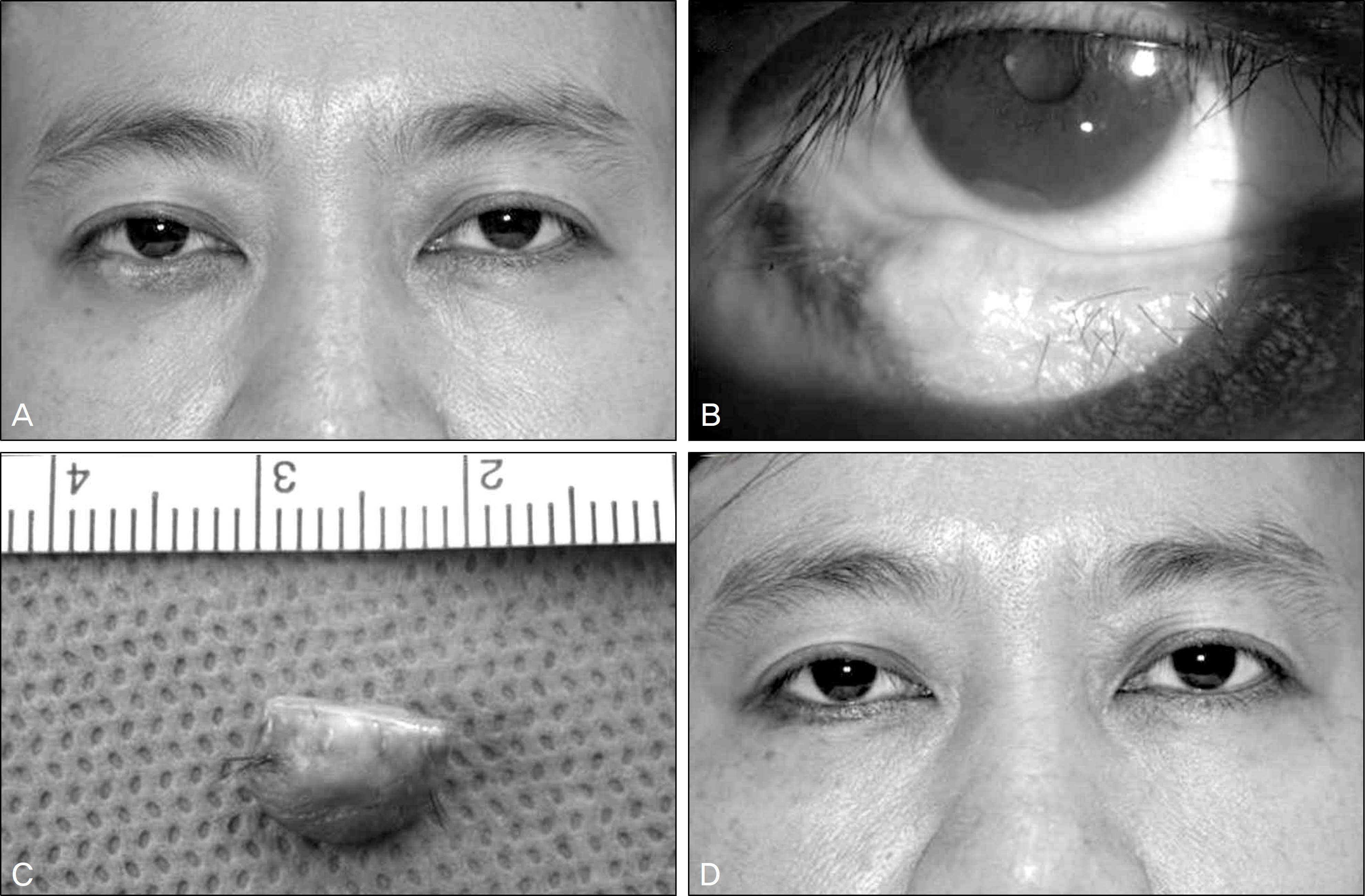
Figure 1. (A) Photograph of the patient demonstrating a 7×7-mm-sized mass in the margin of the right lower eyelid. (B) The mass was pinkish and smooth-surfaced, and the cilia on its surface were lost. (C) Gross photograph of excised eyelid mass. Pentagon-shaped tissue of about 8-mm width, including the eyelid mass and adjacent normal tissue, was excised. (D) Facial photograph taken at postoperative 2 weeks showing good cosmetic results.

Figure 2. (A) Nests and sheets of epithelioid cells dispersed in a collagenous and myxoid stroma (hematoxylin-eosin, ×40). (B) Cuboidal epithelial cells, ductal structures (arrow), and multiple cysts are noted (hematoxylin-eosin, ×200).
Reference
-
References
1. Hirsch P, Helwig EB. Chondroid syringoma. Mixed tumor of skin, salivary gland type. Arch Dermatol. 1961; 84:835–47.2. Kitazawa T, Hataya Y, Matsuo K. Chondroid syringoma of the orbit. Ann Plast Surg. 1999; 42:100–2.
Article3. Yavuzer R, Baş terzi Y, Sari A, et al. Chondroid syringoma: a diagnosis more frequent than expected. Dermatol Surg. 2003; 29:179–81.
Article4. Park CY, Chi JY, Suh YL, Kim YD. Chondroid syringoma of the eyelid. J Korean Ophthalmol Soc. 2003; 44:1684–8.5. Gündüz K, Demirel S, Heper AO, Günalp I. A rare case of atypical chondroid syringoma of the lower eyelid and review of the literature. Surv Ophthalmol. 2006; 51:280–5.
Article6. Jordan DR, Nerad JA, Patrinely JR. Chondroid syringoma of the eyelid. Can J Ophthalmol. 1989; 24:24–7.7. Mandeville JT, Roh JH, Woog JJ, et al. Cutaneous benign mixed tumor (chondroid syringoma) of the eyelid: clinical presentation and management. Ophthal Plast Reconstr Surg. 2004; 20:110–6.
Article8. Martorina M, Capoferri C, Dessanti P. Chondroid syringoma of the eyelid. Int Ophthalmol. 1993; 17:285–8.
Article9. Mencía-Gutiérrez E, Bonales-Daimiel JA, Gutiérrez-Díaz E, et al. Chondroid syringomas of the eyelid: two cases. Eur J Ophthalmol. 2001; 11:80–2.
Article11. Tyagi N, Abdi U, Tyagi SP, et al. Pleomorphic adenoma of skin (chondroid syringoma) involving the eyelid. J Postgrad Med. 1996; 42:125–6.12. Ogawa R, Mitsuhashi K, Oki K, Hyakusoku H. A rare case of chondroid syringoma arising from the lower eyelid with ectropion. Plast Reconstr Surg. 2006; 118:137e–40e.
Article13. Shields JA, Shields CL, Eagle RC Jr. Trichoadenoma of the eyelid. Am J Ophthalmol. 1998; 126:846–8.
Article
