J Korean Ophthalmol Soc.
2011 Jul;52(7):807-815. 10.3341/jkos.2011.52.7.807.
Analysis of Factors Affecting Corneal Endothelial Cell Loss after Penetrating Keratoplasty
- Affiliations
-
- 1Department of Ophthalmology and Visual Science, The Catholic University of Korea School of Medicine, Seoul, Korea. mskim@catholic.ac.kr
- KMID: 2214705
- DOI: http://doi.org/10.3341/jkos.2011.52.7.807
Abstract
- PURPOSE
To evaluate the factors affecting corneal endothelial cell loss after penetrating keratoplasty in a long-term follow-up.
METHODS
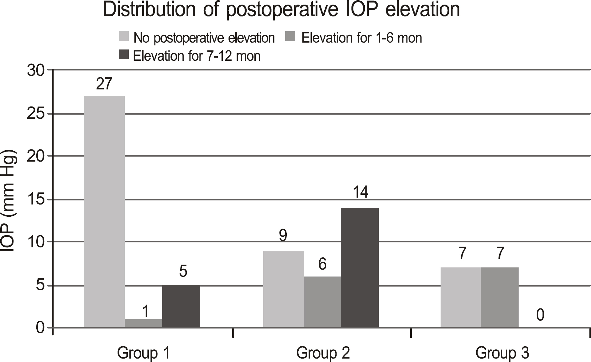
Donor age, post-mortem time, storage time, underlying disease, elevation of IOP after surgery, underlying glaucoma, and trephine size were analyzed in 76 eyes. Postoperative corneal endothelial density was measured after 1, 3, 6, and 12 months. Patients who experienced graft rejection were excluded.
RESULTS
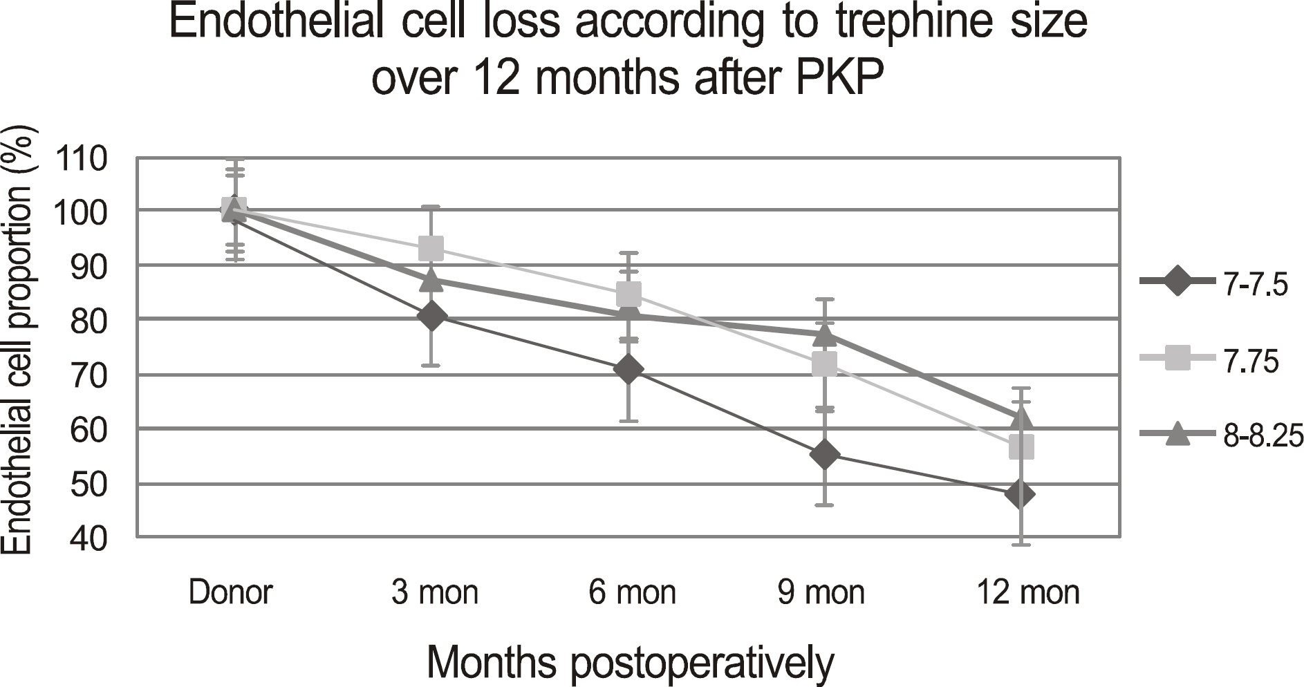
Donor age and endothelial loss were correlated in all patients (t-value = 1.98); however, post-mortem time and storage time were not statistically significant (t 2 < 2). Endothelial cell loss was more severe in the bullous keratopathy patient group than it was in the keratoconus patient group, but this difference was not statistically significant (p-value = 0.154). The number of anti-glaucomatous eye drops showed positive correlation with the declining rate of endothelial cells (t-value = 1.975). Existence of glaucoma diagnosed before surgery did not statistically influence endothelial cell loss. Additionally, in the bullous keratopathy patient group, an inverse correlation between endothelial cell loss and trephine diameter was observed (t-value = -2.859).
CONCLUSIONS
Old donor age, small trephine size in bullous keratopathy, and post-operative IOP elevation are risk factors for increased endothelial cell loss following penetrating keratoplasty.
MeSH Terms
Figure
Cited by 1 articles
-
Influence of Endothelial Cell Loss During Preservation on Graft Survival in Imported Donor Cornea
Kook Lee, Kyu Yeon Hwang, Man Soo Kim
J Korean Ophthalmol Soc. 2013;54(6):862-868. doi: 10.3341/jkos.2013.54.6.862.
Reference
-
References
1. Culbertson WW, Abbott RL, Forster RK. Endothelial cell loss in penetrating keratoplasty. Ophthalmology. 1982; 89:600–4.
Article2. Armitage WJ, Dick AD, Bourne WM. Predicting endothelial cell loss and long-term corneal graft survival. Invest Ophthalmol Vis Sci. 2003; 44:3326–31.
Article3. Wollensak G, Green WR. Analysis of sex-mismatched human corneal transplants by fluorescence in situ hybridization of the sex-chromosomes. Exp Eye Res. 1999; 68:341–6.
Article4. Bell KD, Campbell RJ, Bourne WM. Pathology of late endothelial failure: late endothelial failure of penetrating keratoplasty: study with light and electron microscopy. Cornea. 2000; 19:40–6.
Article5. Bertelmann E, Hartmann C, Scherer M, Rieck P. Outcome of rotational keratoplasty: comparison of endothelial cell loss in autografts vs allografts. Arch Ophthalmol. 2004; 122:1437–40.6. Birnbaum F, Reinhard T, Böhringer D, Sundmacher R. Endothelial cell loss after autologous rotational keratoplasty. Graefes Arch Clin Exp Ophthalmol. 2005; 243:57–9.
Article7. Bertelmann E, Pleyer U, Rieck P. Risk factors for endothelial cell loss postkeratoplasty. Acta Ophthalmol Scand. 2006; 84:766–70.
Article8. Lee HS, Kim MS. Influential factors on the survival of endothelial cells after penetrating keratoplasty. Eur J Ophthalmol. 2009; 19:930–5.
Article9. Langenbucher A, Seitz B, Nguyen NX, Naumann GO. Corneal endothelial cell loss after nonmechanical penetrating keratoplasty depends on diagnosis: a regression analysis. Graefes Arch Clin Exp Ophthalmol. 2002; 240:387–92.
Article10. Chung SH, Kim HK, Kim MS. Corneal endothelial cell loss after penetrating keratoplasty in relation to preoperative recipient endothelial cell density. Ophthalmologica. 2010; 224:194–8.
Article11. Maguire MG, Stark WJ, Gottsch JD, et al. Risk factors for corneal graft failure and rejection in the collaborative corneal transplantation studies. Collaborative Corneal Transplantation Studies Research Group. Ophthalmology. 1994; 101:1536–47.12. Reinhard T, Böhringer D, Sundmacher R. Accelerated chronic endothelial cell loss after penetrating keratoplasty in glaucoma eyes. J Glaucoma. 2001; 10:446–51.
Article13. Nguyen NX, Langenbucher A, Seitz B, et al. Impact of increased intraocular pressure on long-term corneal endothelial cell density after penetrating keratoplasty. Ophthalmologica. 2002; 216:40–4.
Article14. Rho HJ. Multiple regression analysis. Jang JI, editor. Theory and Practice of Multivariate Analysis. 1st ed.Seoul: Hyungseul Publishing Co.;2005. p. 262–303.15. Bourne WM. Chronic endothelial cell loss in transplanted corneas. Cornea. 1983; 2:289–94.
Article16. Bourne WM, Nelson LR, Hodge DO. Central corneal endothelial cell changes over a ten-year period. Invest Ophthalmol Vis Sci. 1997; 38:779–82.17. Bourne WM. Cellular changes in transplanted human corneas. Cornea. 2001; 20:560–9.
Article18. Wagoner MD, Ba-Abbad R, Sutphin JE, Zimmerman MB. Corneal transplant survival after onset of severe endothelial rejection. Ophthalmology. 2007; 114:1630–6.
Article19. Ing JJ, Ing HH, Nelson LR, et al. Ten-year postoperative results of penetrating keratoplasty. Ophthalmology. 1998; 105:1855–65.
Article20. Williams KA, Muehlberg SM, Lewis RF, Coster DJ. Influence of advanced recipient and donor age on the outcome of corneal transplantation. Australian Corneal Graft Registry. Br J Ophthalmol. 1997; 81:835–9.21. Andersen J, Ehlers N. Corneal transplantation using 4-week banked donor material. Long-term results. Acta Ophthalmol. 1987; 65:293–9.
Article22. Andersen J, Ehlers N. The influence of donor age and post mortem time on corneal graft survival and thickness when employing banked donor material (A five-year follow-up). Acta Ophthalmol. 1988; 66:313–7.23. Böhringer D, Reinhard T, Spelsberg H, Sundmacher R. Influencing factors on chronic endothelial cell loss characterised in a homogeneous group of patients. Br J Ophthalmol. 2002; 86:35–8.24. Bourne WM, Nelson LR, Maguire LJ, et al. Comparison of Chen Medium and Optisol-GS for human corneal preservation at 4 degrees C: results of transplantation. Cornea. 2001; 20:683–6.25. Ha DW, Kim CK, Lee SE, et al. Penet rating keratoplasty results in 275 cases. J Korean Ophthalmol Soc. 2001; 42:20–9.26. Kim SH, Ahn BC, Chung YT. Endothelial cell changes after penetrating keratoplasty. J Korean Ophthalmol Soc. 2000; 41:1124–31.27. Patel SV, Hodge DO, Bourne WM. Corneal endothelium and post-operative outcomes 15 years after penetrating keratoplasty. Am J Ophthalmol. 2005; 139:311–9.
Article28. Goldberg DB, Schanzlin DJ, Brown SI. Incidence of increased intraocular pressure after keratoplasty. Am J Ophthalmol. 1981; 92:372–7.
Article29. Charlin R, Polack FM. The effect of elevated intraocular pressure on the endothelium of corneal grafts. Cornea. 1982; 1:241–9.
Article30. Santoul C, Decrouez E, Driot JY, Bonne C. Use of a specular microscope with pachymeter in ocular tolerance studies of eye drops in the rabbit. Evaluation of ocular tolerance of benzalkonium chloride in aqueous solution 0.01% and 0.1%. Lens Eye Toxic Res. 1990; 7:359–69.31. Zimmerman T, Olson R, Waltman S, Kaufman H. Transplant size and elevated intraocular pressure. Postkeratoplasty. Arch Ophthalmol. 1978; 96:2231–3.
- Full Text Links
- Actions
-
Cited
- CITED
-
- Close
- Share
- Similar articles
-
- Endothelial Cell Changes after Penetrating Keratoplasty
- Endothelial Cell Loss in Penetrating Keratoplasty
- Influence of Endothelial Cell Loss During Preservation on Graft Survival in Imported Donor Cornea
- Cataract Extraction after Penetrating Keratoplasty
- The Surgical Result of Phacoemulsification after Penetrating Keratoplasty


