J Korean Ophthalmol Soc.
2011 Jun;52(6):753-758. 10.3341/jkos.2011.52.6.753.
A Case of Nonarteritic Anterior Ischemic Optic Neuropathy Following Acute Angle-Closure Glaucoma
- Affiliations
-
- 1Department of Ophthalmology, Pusan National University College of Medicine, Busan, Korea. alertlee@hanmail.net
- KMID: 2214651
- DOI: http://doi.org/10.3341/jkos.2011.52.6.753
Abstract
- PURPOSE
Nonarteritic anterior ischemic optic neuropathy (NAION) is believed to result from inadequate blood supply to the posterior ciliary arteries. To date, NAION in a patient with acute angle-closure glaucoma (AACG) has been reported in only two studies in the English literature. Thus, the authors report a case of NAION following AACG in a Korean patient.
CASE SUMMARY
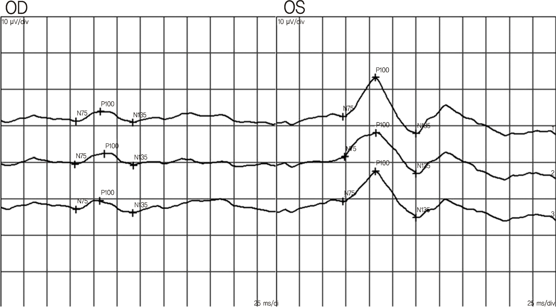
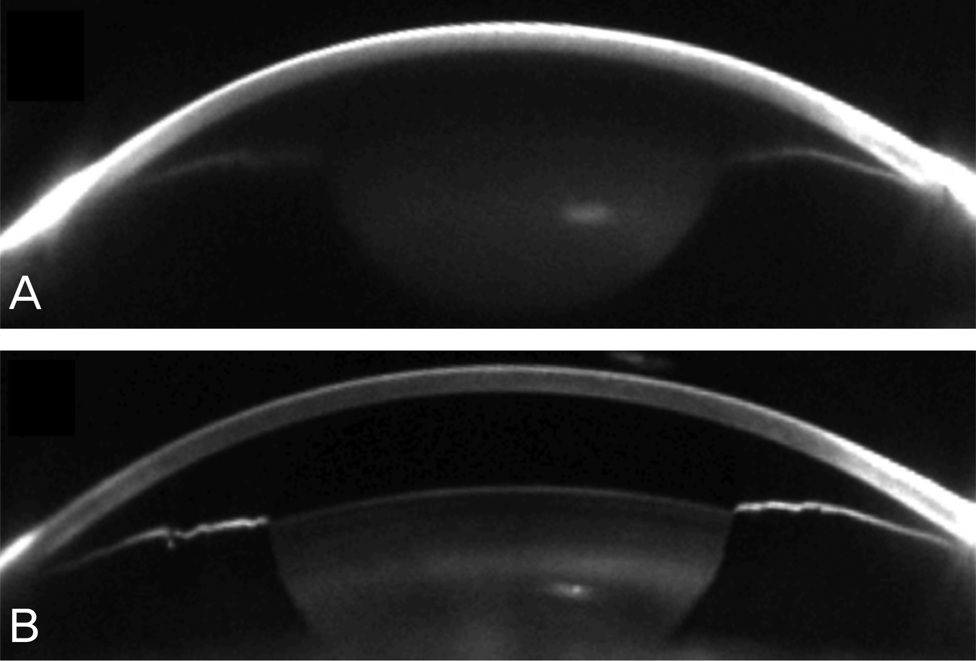
A 59-year-old woman presented with a three-day history of acute ocular pain and decreased vision in her right eye; visual acuity was hand movement and the intraocular pressure (IOP) was 66 mm Hg in the right eye. Slit-lamp examination of the patient's right eye revealed diffuse corneal edema, shallow anterior chamber, and mid-dilated pupil. Gonioscopy revealed a grade 0 angle in the right eye, and a relative afferent pupillary defect was noted. Fundus photography showed disc hemorrhage and swelling of the optic disc. Fluorescein angiography demonstrated hyperfluorescence of the optic disc due to leakage. Visual evoked potential of the right eye at the initial visit showed a decreased amplitude of P100 compared with that of the left eye. A diagnosis of NAION following AACG was made. Laser iridotomy was successfully performed to the right eye. Two months later, IOP decreased from 66 to 21 mm Hg. However, visual acuity remained as hand movement and fundus examination revealed a pale optic disc.
CONCLUSIONS
NAION following AACG may be attributed to an acute IOP rise with resultant perfusion pressure decrease in the vessels which supply the optic nerve. The result obtained from the patient in the present study indicates that evaluation for NAION should be considered in AACG cases.
MeSH Terms
-
Anterior Chamber
Ciliary Arteries
Corneal Edema
Evoked Potentials, Visual
Eye
Female
Fluorescein Angiography
Glaucoma, Angle-Closure
Gonioscopy
Hand
Hemorrhage
Humans
Intraocular Pressure
Middle Aged
Optic Nerve
Optic Neuropathy, Ischemic
Patient Rights
Perfusion
Photography
Pupil
Pupil Disorders
Vision, Ocular
Visual Acuity
Figure
Reference
-
References
1. Jun BK, Kim DS, Ko MK. Clinical features in anterior ischemic optic neuropathy. J Korean Ophthalmol Soc. 1999; 40:3460–7.2. Kim DH, Hwang JM. Risk factors for Korean patients with anterior ischemic optic neuropathy. J Korean Ophthalmol Soc. 2007; 48:1527–31.
Article3. Hayreh SS. Anterior ischemic optic neuropathy. IV. Occurrence after cataract extraction. Arch Ophthalmol. 1980; 98:1410–6.4. Flaharty PM, Sergott RC, Lieb W, et al. Optic nerve sheath decompression may improve blood flow in anterior ischemic optic neuropathy. Ophthalmology. 1993; 100:297–302.
Article5. Slavin ML, Margulis M. Anterior ischemic optic neuropathy following acute angle-closure glaucoma. Arch Ophthalmol. 2001; 119:1215.6. Nahum Y, Newman H, Kurtz S, Rachmiel R. Nonarteritic anterior ischemic optic neuropathy in a patient with primary acute angle-closure glaucoma. Can J Ophthalmol. 2008; 43:723–4.
Article7. Kim R, Van Stavern G, Juzych M. Nonarteritic anterior ischemic optic neuropathy associated with acute glaucoma secondary to Posner-Schlossman syndrome. Arch Ophthalmol. 2003; 121:127–8.
Article8. Lee H, Kim CY, Seong GJ, Ma KT. A case of decreased visual field after uneventful cataract surgery: nonarteritic anterior ischemic optic neuropathy. Korean J Ophthalmol. 2010; 24:57–61.
Article9. Tomsak RL, Remler BF. Anterior ischemic optic neuropathy and increased intraocular pressure. J Clin Neuroophthalmol. 1989; 9:116–8.10. Ho SF, Dhar-Munshi S. Nonarteritic anterior ischaemic optic neuropathy. Curr Opin Ophthalmol. 2008; 19:461–7.
Article11. Doro S, Lessell S. Cup-disc ratio and ischemic optic neuropathy. Arch Ophthalmol. 1985; 103:1143–4.
Article12. McCulley TJ, Lam BL, Feuer WJ. Nonarteritic anterior ischemic optic neuropathy and surgery of the anterior segment: temporal relationship analysis. Am J Ophthalmol. 2003; 136:1171–2.
Article13. Jang BL. Neuroophthalmology. 1st ed.Seoul: Ilchokak;2008. p. 46–58.14. Lan YW, Wang IJ, Hsiao YC, et al. Characteristics of disc hemorrhage in primary angle-closure glaucoma. Ophthalmology. 2008; 115:1328–33.
Article15. Park WC, Chang BL. Clinical features of anterior ischemic optic neuropathy. J Korean Ophthalmol Soc. 2003; 44:144–9.16. Kee HS, Kim SJ, Yang KJ. Clinical study on primary acute angle closure glaucoma. J Korean Ophthalmol Soc. 1995; 36:499–504.17. Arnold AC, Hepler RS. Fluorescein angiography in acute non-arteritic anterior ischemic optic neuropathy. Am J Ophthalmol. 1994; 117:222–30.
Article18. Atilla H, Tekeli O, Ornek K, et al. Pattern electroretinography and visual evoked potentials in optic nerve diseases. Clin Neurosci. 2006; 13:55–9.
Article19. Hayreh SS, Zimmerman MB, Podhajsky P, Alward WL. Nocturnal arterial hypotension and its role in optic nerve head and ocular ischemic disorders. Am J Ophthalmol. 1994; 117:603–24.
Article20. Beck RW, Savino PJ, Repka MX, et al. Optic disc structure in anterior ischemic optic neuropathy. Ophthalmology. 1984; 91:1334–7.
Article21. Hayreh SS, Podhajsky P, Zimmerman MB. Role of nocturnal arterial hypotension in optic nerve head ischemic disorders. Ophthalmologica. 1999; 213:76–96.
Article22. Hayreh SS, Podhajsky PA, Zimmerman B. Nonarteritic anterior ischemic optic neuropathy: time of onset of visual loss. Am J Ophthalmol. 1997; 124:641–7.
Article
- Full Text Links
- Actions
-
Cited
- CITED
-
- Close
- Share
- Similar articles
-
- Nonarteritic Anterior Ischemic Optic Neuropathy Accompanying Appositional Angle-closure Glaucoma Mimicking Glaucomatocyclitic Crisis
- Bilateral Delayed Nonarteritic Anterior Ischemic Neuropathy Following Acute Primary Angle-closure Crisis
- Delayed Non-arteritic Anterior Ischemic Optic Neuropathy Following Acute Primary Angle Closure
- Retinal Nerve Fiber Layer-to-Disc Ratio Distinguishing Glaucoma from Nonarteritic Anterior Ischemic Optic Neuropathy
- The Effect of an Intravitreal Triamcinolone Acetonide Injection for Acute Nonarteritic Anterior Ischemic Optic Neuropathy


