J Korean Ophthalmol Soc.
2011 Jun;52(6):709-715. 10.3341/jkos.2011.52.6.709.
Clinical Validation of Visual Field Index in Glaucoma Patients with Central Visual Field Defects
- Affiliations
-
- 1Department of Ophthalmology, Korea University College of Medicine, Seoul, Korea. yongykim@korea.ac.kr
- KMID: 2214643
- DOI: http://doi.org/10.3341/jkos.2011.52.6.709
Abstract
- PURPOSE
To evaluate the glaucoma discrimination ability of visual field index (VFI), a new perimetric index of Humphrey field analyzer II, in glaucoma patients with central and peripheral visual field defects (VFD).
METHODS
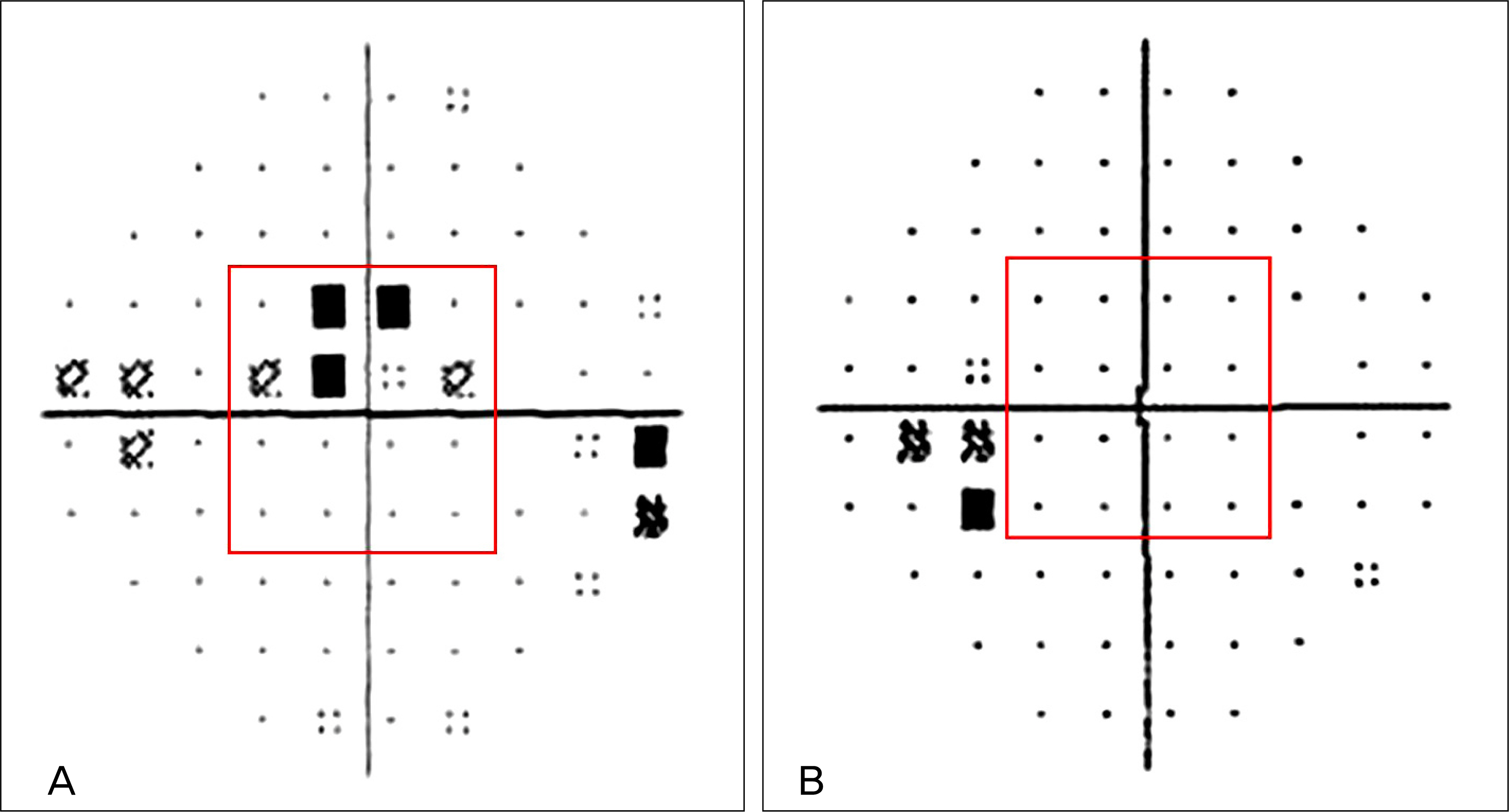
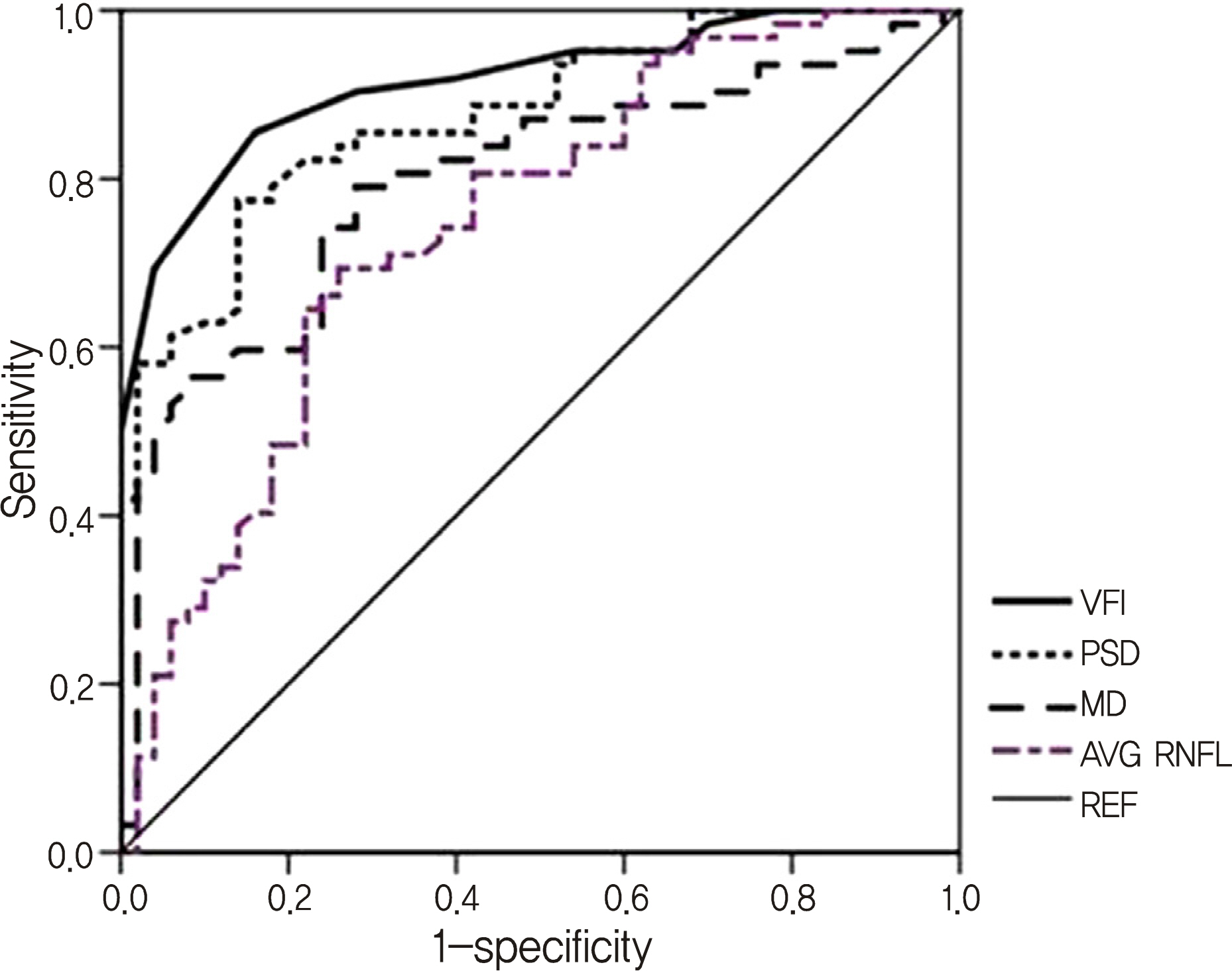
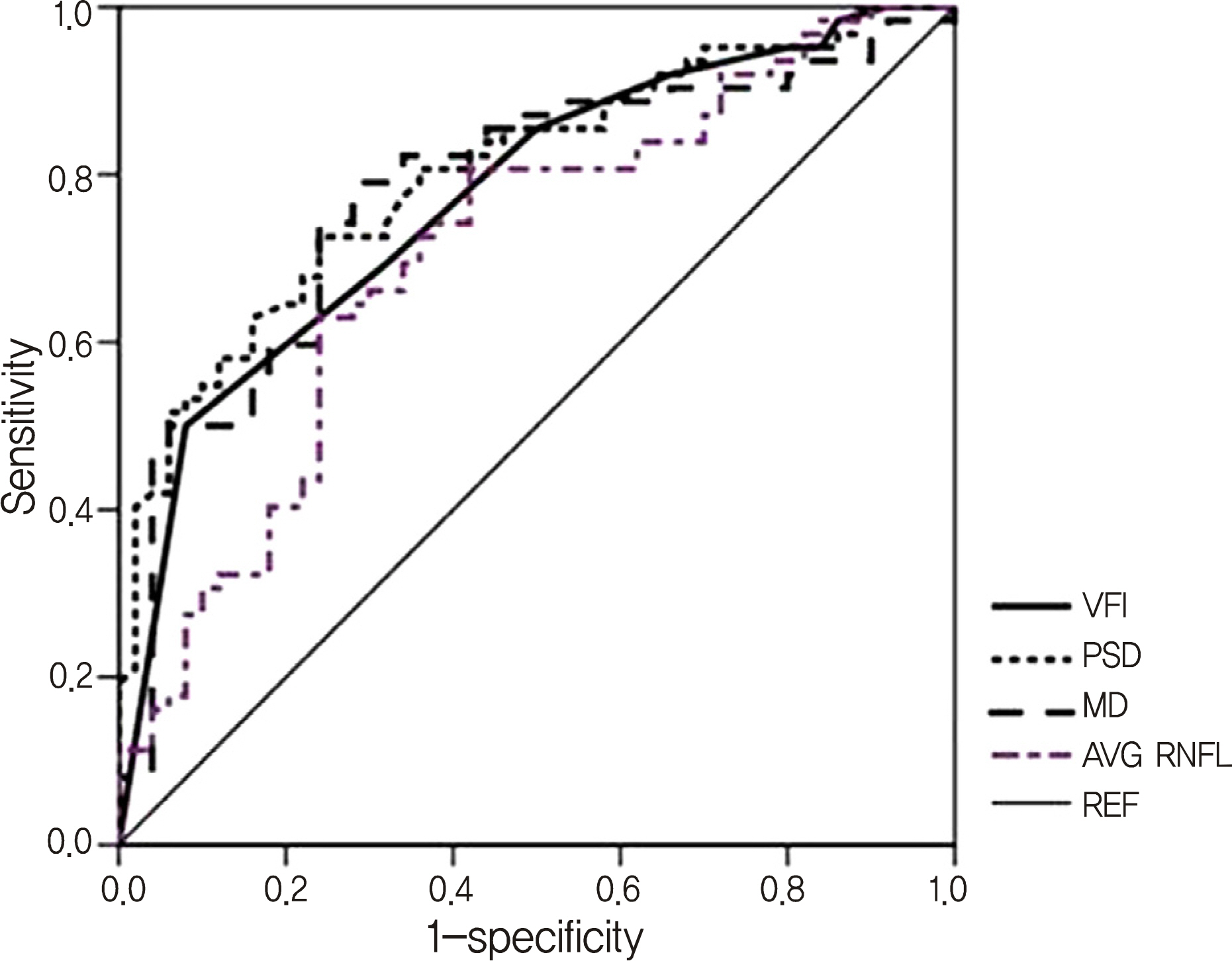
Humphrey visual field test and OCT were performed in 204 glaucomatous eyes and 70 healthy eyes. The associations of VFI with mean deviation (MD), pattern standard deviation (PSD), and average retinal nerve fiber layer thickness (RNFLT) were analyzed using Pearson's correlation. The diagnostic abilities of the parameters were analyzed using the areas under the receiver operating characteristic curves (AUROC). The AUROC were compared between MD-matched patients with central VFD (at least one point with p < 1% within the central most 16 points of 30-2 SITA standard automated visual field) and peripheral VFD (VFD beyond the central most 16 points of 30-2 SITA standard automated visual field).
RESULTS
The associations between analyzed parameters and VFI were statistically significant. The MD, RNFLT, age, intraocular pressure, and central cornea thickness were not different between the two groups (p > 0.05). The AUROC value of VFI was greater than those of the MD and average RNFLT but was not different from that of PSD (p = 0.332) in the central VFD group. However, there were no significant differences between AUROC value of VFI and those of other parameters in the peripheral VFD group (all, p > 0.05).
CONCLUSIONS
The results from the present study suggest that VFI may be more useful than MD in diagnosing glaucoma, especially in patients with central VFD.
Keyword
MeSH Terms
Figure
Reference
-
References
1. Sommer A, Miller NR, Pollack I, et al. The nerve fiber layer in the diagnosis of glaucoma. Arch Ophthalmol. 1977; 95:2149–56.
Article2. Cho CH, Kee CW. Association of retinal nerve fiber layer thickness measured by optical coherence tomography and automatic perimetry. J Korean Ophthalmol Soc. 2002; 43:1032–9.3. Quigley HA, Dunkelberger GR, Green WR. Retinal ganglion cell atrophy correlated with automated perimetry in human eyes with glaucoma. Am J Ophthalmol. 1989; 107:453–64.
Article4. Sommer A, Katz J, Quigley HA, et al. Clinically detectable nerve fiber atrophy precedes the onset of glaucomatous field loss. Arch Ophthalmol. 1991; 109:77–83.
Article5. Heijl A, Lindgren G, Olsson J. A package for the statistical analy-sis of visual fields. Doc Ophthalmol Proc Ser. 1987; 49:153–68.
Article6. Bengtsson B, Heijl A. A visual field index for calculation of glaucoma rate of progression. Am J Ophthalmol. 2008; 145:343–53.
Article7. Lima VC, Prata TS, De Moraes CG, et al. A comparison between microperimetry and standard achromatic perimetry of the central visual field in eyes with glaucomatous paracentral visual-field defects. Br J Ophthalmol. 2010; 94:64–7.
Article8. Flammer J. Fluctuations in the visual fields. Drance SM, Anderson DR, editors. Automatic Perimetry in Glaucoma. A Practical Guide, New ed. Orlando, FL: Grune and Stratton Inc.;1985. 1:chap. 7.9. Asman P, Heijl A. Diffuse visual field loss and glaucoma. Acta Ophthalmol. 1994; 72:303–8.10. Chauhan BC, LeBlanc RP, Shaw AM, et al. Repeatable diffuse visual field loss in open-angle glaucoma. Ophthalmology. 1997; 104:532–8.
Article11. Weber JT. Topographie der Funktionellen Schädigung Beim Chronishen Glaukom, New ed. Heidelberg, Germany: Kauden Verlag. 1992; 81.12. Asman P, Wild JM, Heijl A. Appearance of the pattern deviation map as a function of change in area of localized field loss. Invest Ophthalmol Vis Sci. 2004; 45:3099–106.13. Artes PH, Nicolela MT, LeBlanc RP, Chauhan BC. Visual field progression in glaucoma: total versus pattern deviation analyses. Invest Ophthalmol Vis Sci. 2005; 46:4600–6.
Article14. Cho JW, Nam YP, Kim DY, et al. Clinical validation of visual field index. J Korean Ophthalmol Soc. 2010; 51:49–54.
Article
- Full Text Links
- Actions
-
Cited
- CITED
-
- Close
- Share
- Similar articles
-
- Analysis of Factors Related of Location of Initial Visual Field Defect in Normal Tension Glaucoma
- Visual Field Defects and Disc Findings in Glaucoma
- Path to Diagnosis and Clinical Characteristics of Advanced Glaucoma at Initial Diagnosis: a Tertiary Single Center Experience
- The Differences of Visual Field Defects in Three Types of Primary Glaucoma
- Long Term Fluctuation of Visual Field in Glaucoma Patients


