J Korean Ophthalmol Soc.
2011 May;52(5):589-596. 10.3341/jkos.2011.52.5.589.
Effect of Amniotic Membrane on Epithelial Thickness and Formation of Hemidesmosomes after Corneal Stromal Wound
- Affiliations
-
- 1Department of Ophthalmology, Chungnam National University College of Medicine, Daejeon, Korea. shchoi@cnu.ac.kr
- KMID: 2214579
- DOI: http://doi.org/10.3341/jkos.2011.52.5.589
Abstract
- PURPOSE
To investigate the effects of an amniotic membrane patch on corneal epithelial thickness and formation of hemidesmosomes during corneal stromal wound healing.
METHODS
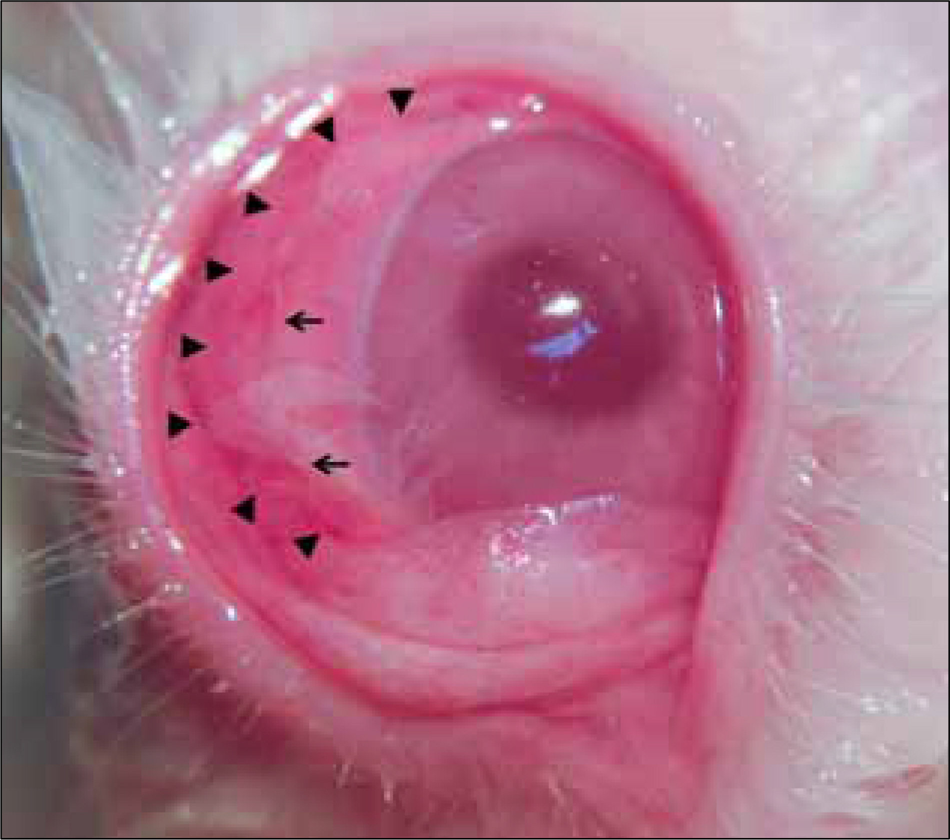
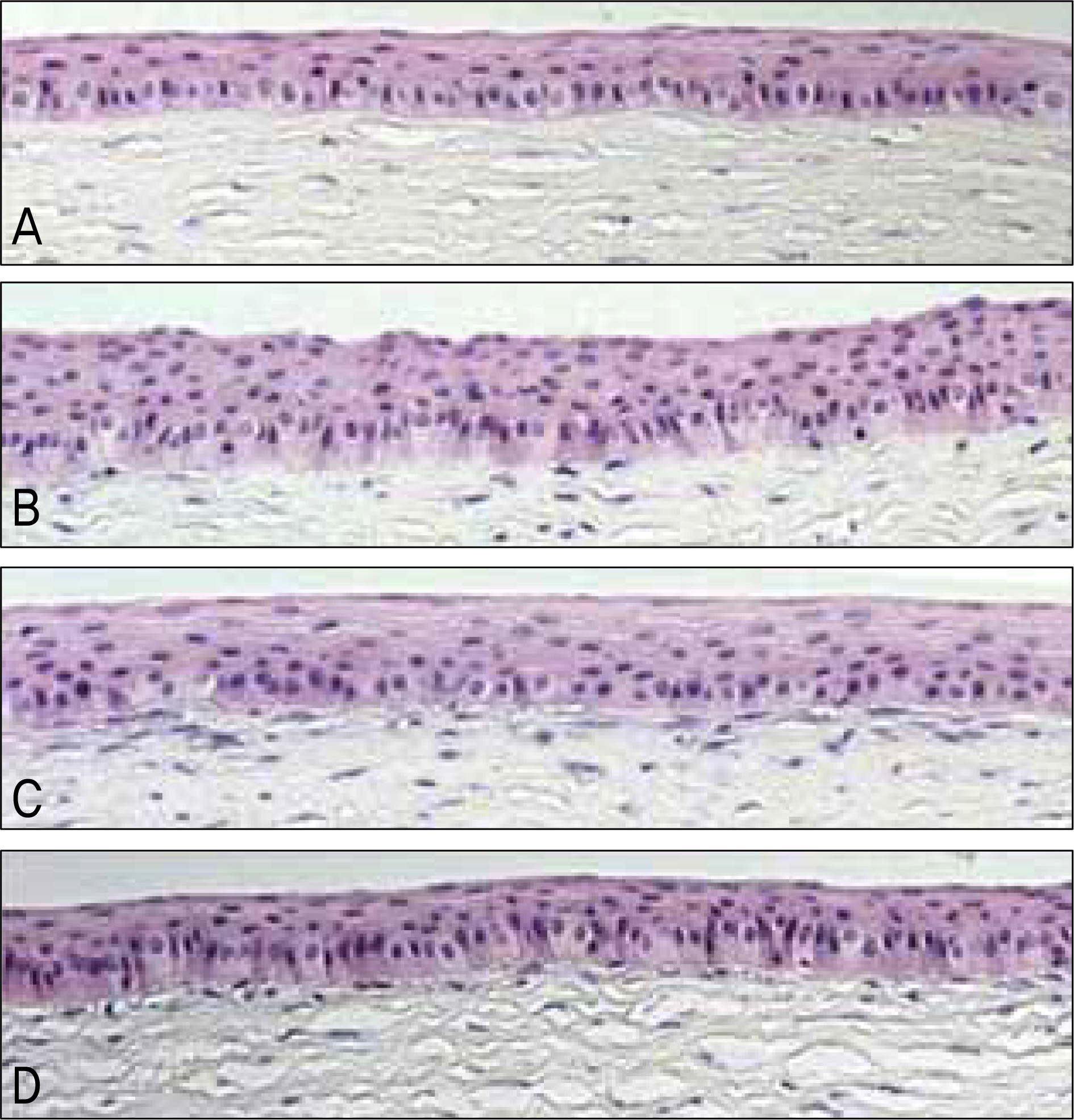
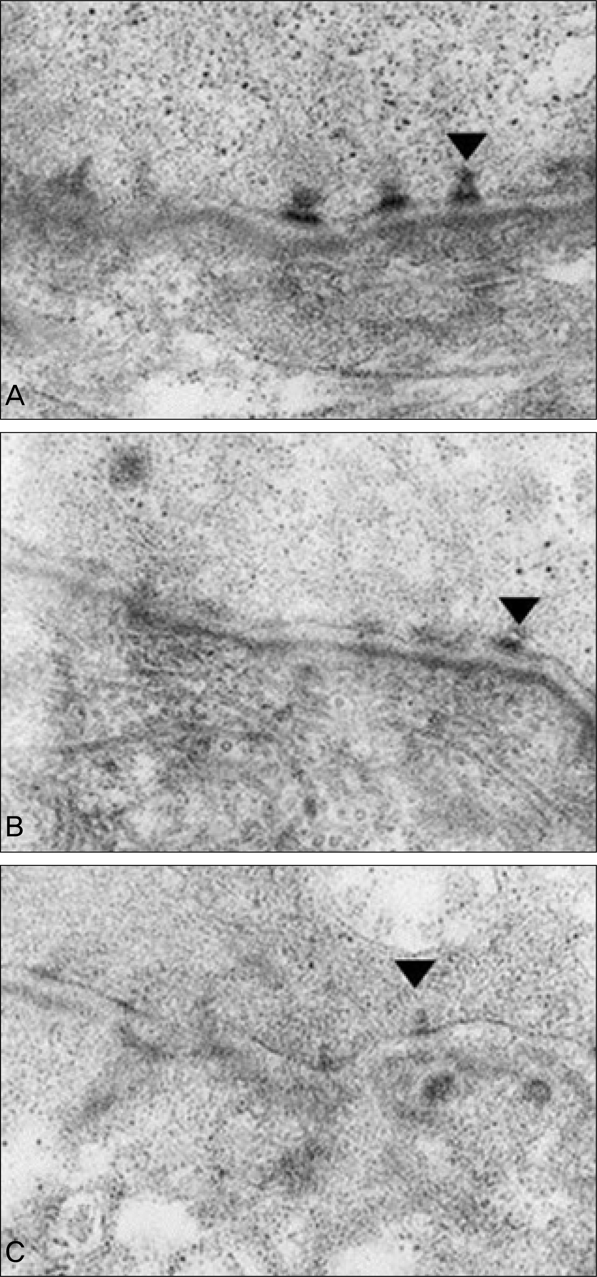
A stromal wound 9 mm in diameter and 130 microm in depth was created on rabbit cornea using a microkeratome. The changes in corneal epithelial thickness and hemidesmosome formations were compared between the amniotic membrane, contact lens, and control groups. Changes in the corneal epithelium were examined using H&E staining and hemidesmosome formation was examined using an electron microscope at 2 and 4 weeks after flap removal.
RESULTS
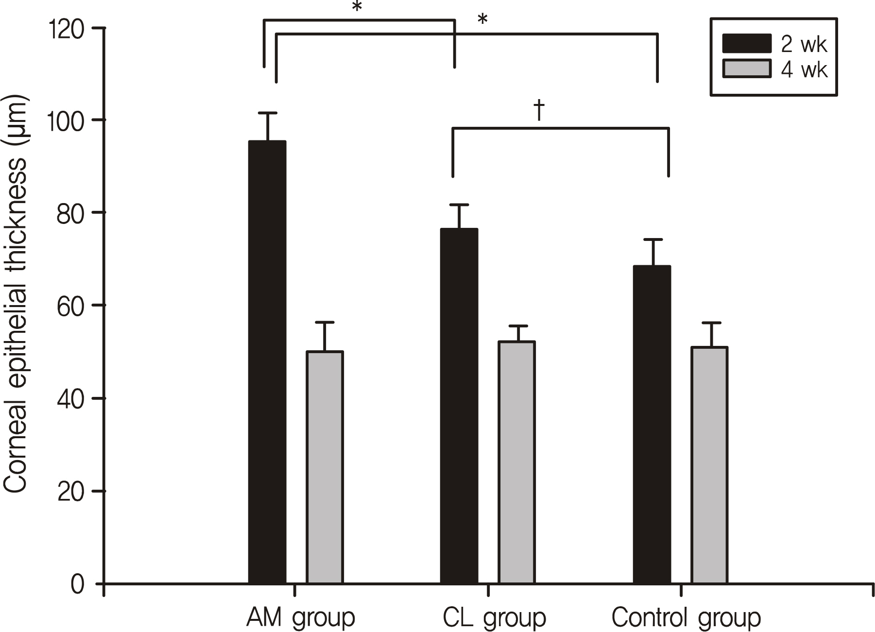
Two weeks after treatment, the corneal epithelial thickness was 95.3 +/- 6.3 microm in the amniotic membrane group being significantly thicker than 76.4 +/- 5.1 microm in the contact lens group and 68.3 +/- 6.1 microm in the control group. Furthermore, more hemidesmosome formations were observed in the amniotic membrane group compared to the other 2 groups. However, there were no significant differences in corneal epithelial thickness or hemidesmosome formation among the 3 groups at week 4.
CONCLUSIONS
The amniotic membrane group showed a thicker corneal epithelium and more hemidesmosome formation than the other 2 groups 2 weeks after flap removal. Thus, the use of an amniotic membrane patch appears to be effective in the early stages of corneal stromal wound healing.
Figure
Reference
-
References
1. Davis JW. Skin transplantation with a review of 550 cases at the Johns Hopkins Hospital. Johns Hopkins Med J. 1910; 15:307.2. Kim JC, Tseng SC. Transplantation of preserved human amniotic membrane for surface reconstruction in severely damaged rabbit corneas. Cornea. 1995; 14:473–84.
Article3. Meller D, Pires RT, Mack RJ, et al. Amniotic membrane transplantation for acute chemical or thermal burns. Ophthalmology. 2000; 107:980–9.
Article4. Kruse FE, Rohrschneider K, Völcker HE. Multilayer amniotic membrane transplantation for reconstruction of deep corneal ulcers. Ophthalmology. 1999; 106:1504–10.
Article5. Chen HJ, Pires RT, Tseng SC. Amniotic membrane transplantation for severe neurotrophic corneal ulcers. Br J Ophthalmol. 2000; 84:826–33.
Article6. Kim JS, Kim JC, Hahn TW, Park WC. Amniotic membrane transplantation in infectious corneal ulcer. Cornea. 2001; 20:720–6.
Article7. Solomon A, Pires RT, Tseng SC. Amniotic membrane transplantation after extensive removal of primary and recurrent pterygia. Ophthalmology. 2001; 108:449–60.
Article8. Hanada K, Shimazaki J, Shimmura S, Tsubota K. Multilayered amniotic membrane transplantation for severe ulceration of the cornea and sclera. Am J Ophthalmol. 2001; 131:324–31.
Article9. van Herendael BJ, Oberti C, Brosens I. Microanatomy of the human amniotic membranes. A light microscopic, transmission, and scanning electron microscopic study. Am J Obstet Gynecol. 1978; 131:872–80.10. Dua HS, Azuara-Blanco A. Amniotic membrane transplantation. Br J Ophthalmol. 1999; 83:748–52.11. Kim JC, Tseng SC. The effects on inhibition of corneal neo-vascularization after human amniotic membrane transplantation in severely damaged rabbit corneas. Korean J Ophthalmol. 1995; 9:32–46.
Article12. Kim SY. Korea External Rye Disease Society. Cornea. 2nd ed.Seoul: Ilchokak;2005. p. 2–3.13. Kim YH, Lee DH, Lew HM. The comparison of the corneal epithelial healing according to treatment modalities in rabbit. J Korean Ophthalmol Soc. 1999; 40:2683–92.14. Kang KM, Wee WR. The use if ascorbic acid after excimer laser photorefractive keratectomy in rabbits. J Korean Ophthalmol Soc. 1996; 37:1620–5.15. Sohn JH, Choi SK, Lee JH. The effect of disposable bandage contact lenses on time and velocity of corneal epithelial healing after myopic epikeratoplasty. J Korean Ophthalmol Soc. 1995; 36:1422–8.16. Fernandes M, Sridhar MS, Sangwan VS, Rao GN. Amniotic membrane transplantation for ocular surface reconstruction. Cornea. 2005; 24:643–53.
Article17. Li H, Niederkorn JY, Neelam S, et al. Immunosuppressive factors secreted by human amniotic epithelial cells. Invest Ophthalmol Vis Sci. 2005; 46:900–7.
Article18. Cooper LJ, Kinoshita S, German M, et al. An investigation into the composition of amniotic membrane used for ocular surface reconstruction. Cornea. 2005; 24:722–9.
Article19. Gomes JA, Romano A, Santos MS, Dua HS. Amniotic membrane use in ophthalmology. Curr Opin Ophthalmol. 2005; 16:233–40.
Article20. Wilson SE, He YG, Weng J, et al. Epithelial injury induces keratocyte apoptosis: hypothesized role for the interleukin-1 system in the modulation of corneal tissue organization and wound healing. Exp Eye Res. 1996; 62:325–7.
Article21. Tseng SC, Li DQ, Ma X. Suppression of transforming growth factor-beta isoforms, TGF-beta receptor type II, and myofibroblast differentiation in cultured human corneal and limbal fibroblasts by amniotic membrane matrix. J Cell Physiol. 1999; 179:325–35.22. Kim JC. A Practical Guide to Ocular Surface Disorders. 1st ed.Seoul: Naeweh Haksool;2006. p. 20–3.23. Woo HM, Kim HS, Kweon OK, et al. Effects of amniotic membrane on epithelial wound healing and stromal remodeling after excimer laser keratectomy in rabbit cornea. Br J Ophthalmol. 2001; 85:345–9.24. Yoshita T, Kobayashi A, Sugiyama K, Tseng SC. Oxygen permeability of amniotic membrane and actual tear oxygen tension beneath amniotic membrane patch. Am J Ophthalmol. 2004; 138:486–7.
Article25. Zagon IS, Sassani JW, Ruth TB, McLaughlin PJ. Epithelial adhesion complexes and organ culture of the human cornea. Brain Res. 2001; 900:205–13.
Article26. Xu YG, Choi SH, Ko SM, et al. Morphology and adhesion complex of cultured epithelium, on amniotic membrane in vitro and in vivo. J Korean Ophthalmol Soc. 2006; 47:160–70.
- Full Text Links
- Actions
-
Cited
- CITED
-
- Close
- Share
- Similar articles
-
- The Changes of Marginal Epithelial Cells in Corneal Wound Healing
- Therapeutic Effect of Amniotic Membrane Extract on Keratitis Following Corneal Alkali Burn
- The Effect of Amniotic Membrane Graft on the Inhibition of Corneal Opacity After Traumatic Injury
- The Effect of Amniotic Membrane Gra ft on the Inhibition of Corneal Haze in Rabbit
- Effect of Topical Na-Hyaluronan on Epithelial Cell Morphogenesis and Superficial Stromal Healing in Corneal Wound


