J Korean Ophthalmol Soc.
2011 Jan;52(1):1-6. 10.3341/jkos.2011.52.1.1.
Comparison of Dacryocystographic Results Before and After Silicone Intubation in Incomplete Nasolacrimal Duct Obstruction
- Affiliations
-
- 1Department of Ophthalmology, Chonbuk National University Medical School, Jeonju, Korea. ahnmin@jbnu.ac.kr
- KMID: 2214130
- DOI: http://doi.org/10.3341/jkos.2011.52.1.1
Abstract
- PURPOSE
To compare the dacryocystographic results before and after silicone tube intubation in partial nasolacrimal duct obstruction.
METHODS
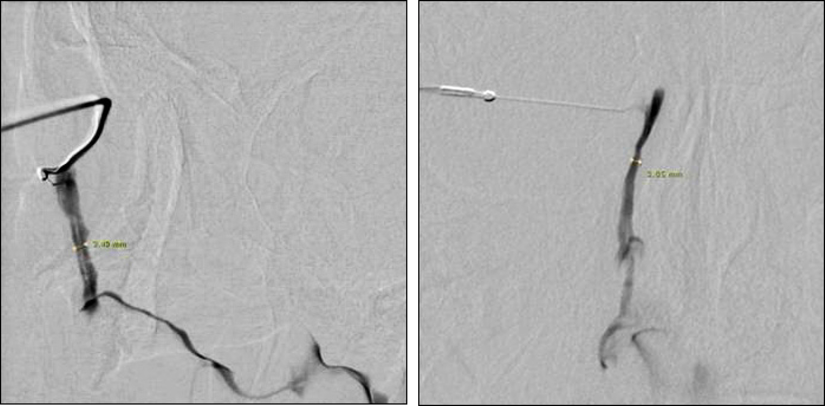
Dacryocystography was performed on 33 eyes of 17 patients diagnosed with partial nasolacrimal duct obstruction. The anteroposterior (AP) diameters and the mediolateral diameters of the nasolacrimal ducts intubated at the operation were measured by dacryocystography, before the operation and after silicone tube removal.
RESULTS
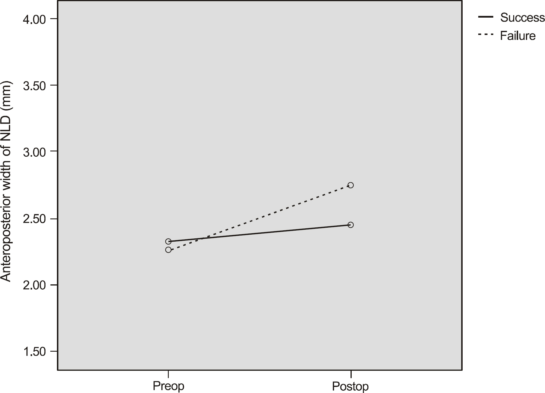
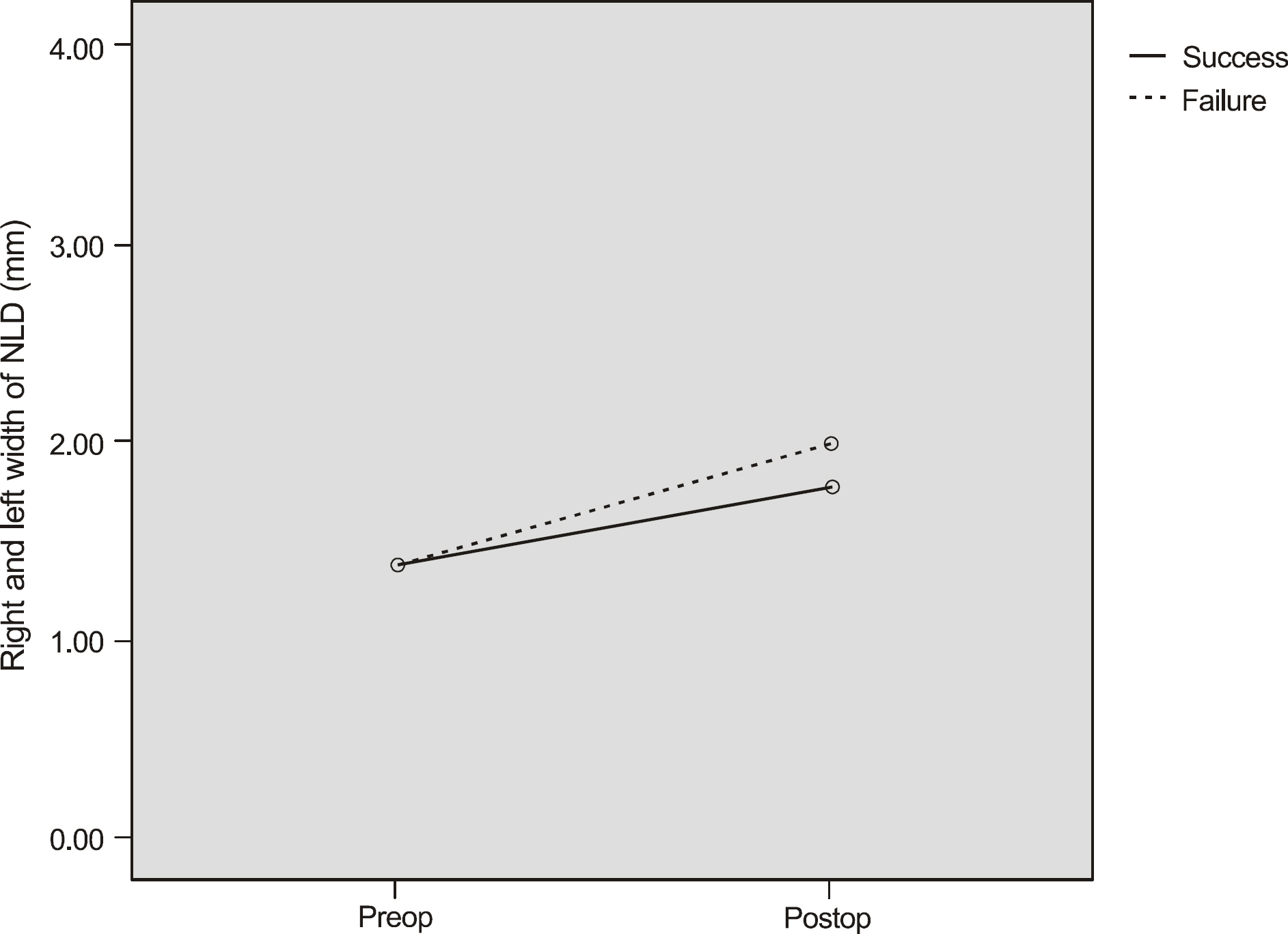
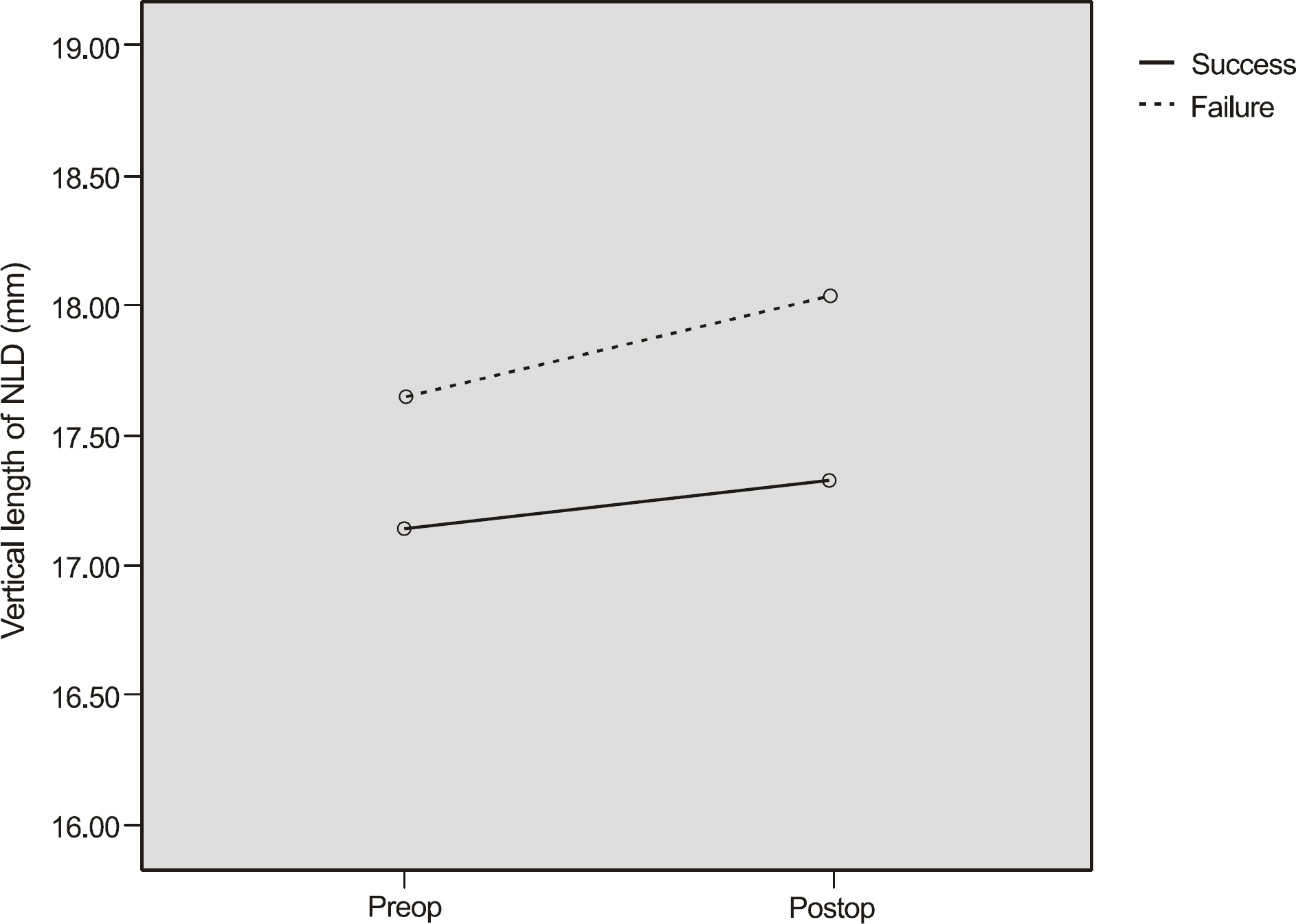
The mean AP, mediolateral diameter and length of nasolacrimal duct in the group who demonstrated improvement after the operation was 2.32 mm, 1.39 mm, and 17.14 mm before the operation, and 2.40 mm, 1.77 mm, and 17.38 mm after the operation, respectively. The mean AP, mediolateral diameter and length of nasolacrimal duct in the group who demonstrated no symptomatic improvement was 2.06 mm, 1.28 mm, and 17.42 mm before the operation, and 2.75 mm, 1.99 mm, and 18.03 mm after the operation, respectively. The alteration of the nasolacrimal duct size in the group with successful postoperative results compared with unsuccessful postoperative results showed no significant difference.
CONCLUSIONS
The nasolacrimal duct showed expansion in size based on dacryocystographic results after silicone tube intubation in partial nasolacrimal duct obstruction. However, the operation results and the alteration of the nasolacrimal duct size based on dacryocystographic results demonstrated no accordance.
Figure
Reference
-
References
1. Jones LT. An anatomical approach to problems of the eyelids and lacrimal apparatus. Arch Ophthalmol. 1961; 66:111–24.
Article2. Kanski JJ. Disorders of the lacrimal drainage system. Clinical ophthalmology. 1999; 43–52.3. Jeffrey JH, Myron Y, Jay SD. The lacrimal drainage system. Ophthalmology. 1999; 7:71–8.4. Ewing AE. Roentgen ray demonstration of the lacrimal abscess cavity. Am J Ophthalomol. 1909; 26:1–4.5. Milder B, Demorest BH. Dacryocystography. I. the normal lacrimal apparatus. AMA Arch Ophthalmol. 1954; 51:180–95.6. Nixon J, Birchall IW, Virjee J. The role of dacryocystography in the management of patient with epiphora. Br J Radiol. 1990; 63:337–9.7. Hurwitz JJ, Welham RA, Lloyd GA. The role of intubation mac-ro-dacryocystography in management of problems of the lacrimal system. Can J Ophthalmol. 1975; 10:361–6.8. Oum JS, Park JW, Choi YK, et al. Result of partial nasolacrimal duct obstruction after silicone tube intubation. J Korean Ophthalmol Soc. 2004; 45:1777–82.9. Campbell W. The radiology of the lacrimal system. Br J Radiol. 1964; 37:1–26.
Article10. Jeong HW, Cho NC, Ahn M. Result of silicone tube intubation in patients with epiphora who showing normal finding in dacryocystography. J Korean Ophthalmol Soc. 2008; 49:706–12.
Article11. Kim DM, Roh KK. Results with silicone stent in lacrimal drainage system. J Korean Ophthalmol Soc. 1987; 28:733–5.12. Sohn HY, Hur J, Chung EH, Won IG. Clinical observation on silicone intubation in obstruction of lacrimal drainage system. J Korean Ophthalmol Soc. 1990; 31:135–40.13. Lee SH, Kim SD, Kim JD. Silicone intubation for nasolacrimal duct obstruction in adult. J Korean Ophthalmol Soc. 1997; 38:185–9.14. Lee HS, Hwang WS, Byun YJ. Clinical results of silicone intubation for nasolacrimal duct obstruction in adult. J Korean Ophthalmol Soc. 1997; 38:1926–30.15. Kim HD, Jeong SK. Silicone tube intubation in acquired nasolacrimal duct obstruction. J Korean Ophthalmol Soc. 2000; 41:327–31.16. Park HJ, Hwang WS. Clinical results of silicone intubation for epiphora patients. J Korean Ophthalmol Soc. 2000; 41:2327–31.17. Lee HS, Lew HL, Yun YS. Classification of nasolacrimal duct obstruction according to dacryocystographic finding and its clinical significance. J Korean Ophthalmol Soc. 2003; 44:1475–82.18. Suh SC, Ha MS. Silicone intubation and dacryocytographic finding in incomplete nasolacrimal duct obstruction. J Korean Ophthalmol Soc. 2009; 50:491–6.19. Lee SU, Kim EH, Lee JE, et al. The clinical outcome of silicone tube intubation according to nasolacrimal duct obstruction sites by dacryoscintigraphy. J Korean Ophthalmol Soc. 2006; 47:863–70.20. Angrist RC, Dortzbach RK. Silicone intubation for partial and total nasolacrimal duct obstruction in adults. Ophthal Plast Reconstr Surg. 1985; 1:51–4.
Article21. Crawford JS. Intubation of the lacrimal system.Ophthal Plast Reconstr Surg. 1989; 5:261–5.22. Chung WS, Park NG. Functional obstruction of the lacrimal drainage system. J Korean Ophthalmol Soc. 1995; 36:1435–8.23. Ruby AJ, Lissner GS, O' Grady R. Surface reaction on silicone tubes used in the treatment of nasolacrimal drainage system obstruction. Ophthalmic Surg. 1991; 22:745–8.
Article
- Full Text Links
- Actions
-
Cited
- CITED
-
- Close
- Share
- Similar articles
-
- Success Rate of Silicone Intubation Between Nasolacrimal Duct Obstruction and Stenosis According to Dacryocystography
- Silicone Intubation and Dacryocystographic Finding in Incomplete Nasolacrimal Duct Obstruction
- Classification of Nasolacrimal Duct Obstruction According to Dacryocystographic Finding and Its Clinical Ssignificance
- Clinical Results of silicone Intubation for Nasolacrimal Duct Obstruction in Adult
- Silicone Intubation for Nasolacrimal Duct Obstruction in Adult


