Surgical Repair of Canalicular Defects and Congenital Eyelid Colobomas Associated with Tessier No. 3 Cleft
- Affiliations
-
- 1Department of Ophthalmology, Seoul National University College of Medicine, Seoul Artificial Eye Center, Seoul National University Hospital Clinical Research Institute, Seoul, Korea. resourceful@hanmail.net
- 2Department of Ophthalmology, Seoul National University Bundang Hospital, Seongnam, Korea.
- 3Department of Ophthalmology, Seoul National University Boramae Hospital, Seoul, Korea.
- KMID: 2213989
- DOI: http://doi.org/10.3341/jkos.2010.51.11.1520
Abstract
- PURPOSE
To report a single case of surgical repair of the canalicular defects and congenital eyelid colobomas associated with Tessier No. 3 craniofacial cleft.
CASE SUMMARY
A one-month-old girl presented with eyelid colobomas and discharges from the eyes. The patient was diagnosed with a Tessier No. 3 craniofacial cleft with bilateral lower eyelid colobomas medial to the puncta. At the age of 55 months, examination under general anesthesia revealed mid-canalicular obstructions in both lower canaliculi. After pentagonal excision of eyelid colobomas in the left upper and both lower eyelids, both ends of the canaliculi were found at the cut edge of the lower eyelids. After the repair of canalicular defects and bilateral nasolacrimal duct silicone tube intubation, the primary closure of the eyelid defect was performed layer by layer. Although there was no subjective improvement of epiphora in the left eye, a subjective improvement of epiphora in the right eye was achieved, and tear meniscus height in the right eye was halved. Additionally, the eyelid colobomas were cosmetically well repaired at postoperative 6 weeks. The patient still had mild tearing symptoms, but did not complain any longer of discharge at postoperative 4 months.
CONCLUSIONS
Tessier No. 3 craniofacial cleft with eyelid colobomas can be associated with canalicular defects and nasolacrimal duct obstructions. Surgical repair of the canalicular defects associated with eyelid colobomas should be considered to achieve a functional recovery of the lacrimal drainage system.
MeSH Terms
Figure
-
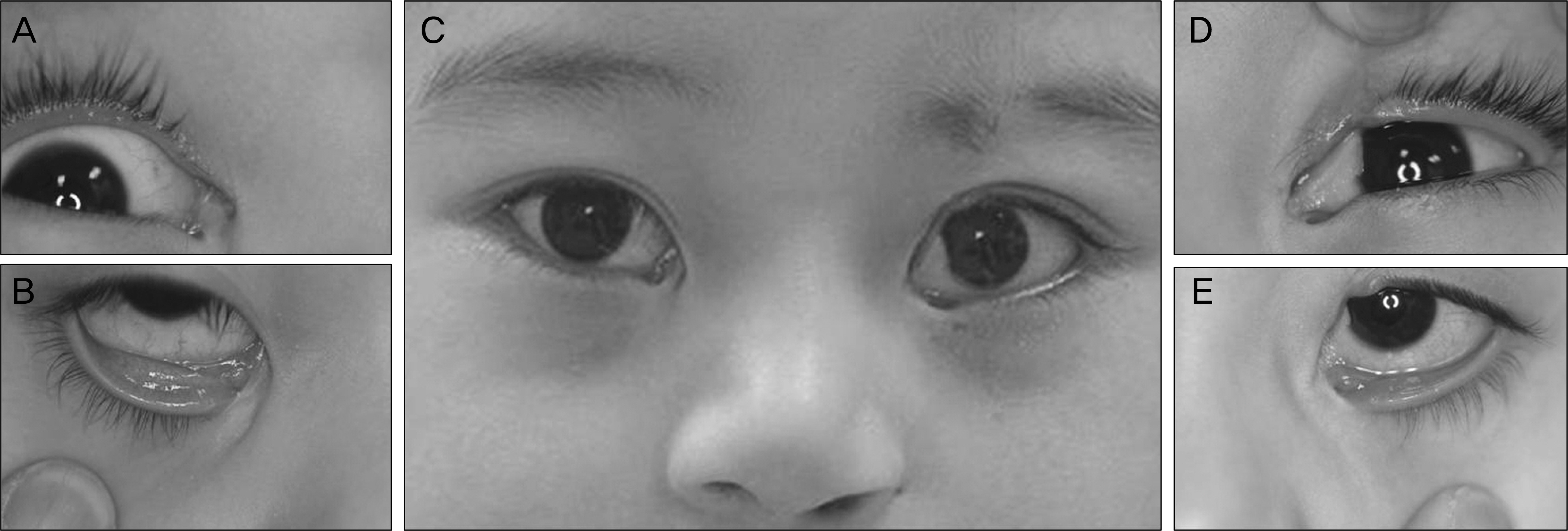
Figure 1. Photographs of the patient before surgery. (A) Intact right upper eyelid. (B) Right lower eyelid coloboma medial to the punctum (C) Preoperative eyelid photograph. Left eyebrow defect. (D) Left upper eyelid coloboma lateral to the punctum, and symblepharon between the left medial canthus and medial conjunctiva adjacent to the limbus. (E) Left lower eyelid coloboma medial to the punctum.

Figure 2. Photographs of the patient after surgery. (A) Two weeks after surgery. Eyelid colobomas were well reconstructed. The silicone tubes were well positioned and tear meniscus heights were high in both eyes. (B) Six weeks after surgery. Right tear meniscus height was decreased. (C) Four months after surgery. Tear meniscus heights were decreased and eye discharge completely resolved.
Reference
-
References
1. Haik HM, Bullock JD. Eyelid coloboma. Roy FH, editor. Master Techniques in Ophthalmic Surgery. Baltimore: Williams & Wilkins;1995. p. 404–8.2. Gribor P. Surgical repair of congenital colobomas. Trans sect Ophthalmol Am Acad Ophthalmol Otolaryngol. 1975; 79:671–8.3. Mustarde JC. Colobomas of the eyelids. Mustarde JC, editor. Repair and Reconstruction in the Orbital Region. New York: Churchill Livingstone;1980. p. 364–72.4. Tranos L. Mandibulofacial dysostosis associated with dermolipo-ma of the conjuctiva. Am J Ophthalmol. 1954; 37:354–9.5. Kawamoto HK Jr. The kaleidoscopic world of rare craniofacial clefts: order out of chaos (Tessier classification). Clin Plast Surg. 1976; 3:529–72.6. Tessier P. Anatomical classification of facial, craniofacial and lat-ero-facial clefts. J Maxillofac Surg. 1976; 4:69–92.
Article7. Moon JW, Hwang JM. Congenital symblepharon associated with No. 3 craniofacial cleft. J Korean Ophthalmol Soc. 2006; 47:171–4.8. Kim S, Hwang JM. Characteristic ocular findings in patients with craniofacial cleft. Graefes Arch Clin Exp Ophthalmol. 2005; 243:490–2.
Article9. Kinsey JA, Streeten BW. Ocular abnormalities in the median cleft face syndrome. Am J Ophthalmol. 1977; 83:261–6.
Article10. Seah LL, Choo CT, Fong KS. Congenital upper lid colobomas: management and visual outcome. Ophthal Plast Reconstr Surg. 2002; 18:190–5.11. Dufresne CR, Jelks GW. Classification of craniofacial malformations. Smith BC, Nesi FA, Lisman RD, Levine MR, editors. Ophthalmic Plastic & Reconstructive Surgery. St. Louis: Mosby;1998. chap. 51.
Article12. Kidwell ED, Tenzel RR. Repair of congenital colobomas of the lids. Arch Ophthalmol. 1979; 97:1931–2.
Article13. Grover AK, Chaudhuri Z, Malik S, et al. Congenital eyelid colobomas in 51 patients. J Pediatr Ophthalmol Strabismus. 2009; 46:151–9.
Article
- Full Text Links
-
- Actions
-
Cited
- CITED
-
- Close
- Share
-
- Similar articles
-
- Anatomical repair of a bilateral Tessier No. 3 cleft by midfacial advancement
- Soft tissue reconstruction in wide Tessier number 3 cleft using the straight-line advanced release technique
- A comprehensive review of surgical techniques in unilateral cleft lip repair
- Three Cases of Typical Ocular Colobomas
- A Wedge-Shaped Anterior Hairline Extension Associated with a Tessier Number 10 Cleft
