J Korean Ophthalmol Soc.
2016 Apr;57(4):657-661. 10.3341/jkos.2016.57.4.657.
A Case of Recurrent External Ophthalmomyiasis Caused by Lucilia sericata Meigen in a Healthy Patient
- Affiliations
-
- 1Department of Ophthalmology, Chonnam National University Medical School, Gwangju, Korea. wchoi82@hanmail.net
- KMID: 2212802
- DOI: http://doi.org/10.3341/jkos.2016.57.4.657
Abstract
- PURPOSE
To report a case of recurrent external ophthalmomyiasis caused by Lucilia sericata Meigen in a healthy patient.
CASE SUMMARY
A 72-year-old male, who was diagnosed 7 months earlier with ophthalmomyiasis and obtained permanent relief by surgical treatment, presented with severe tearing, redness, foreign body sensation, pain in both eyes and aural discomfort in the left ear. On presentation, visual acuity in both eyes was hand movement. On slit lamp examination, total corneal epithelial defect with stromal infiltrations was observed. However, there were no motile organisms in his eyes due to previous removal of maggots at a local clinic. On physical examination, eschar change was observed at the inferomedial area of the left lower eyelid. On otorhinolaryngological examination, several motile organisms with tympanic membrane perforation were observed in the left external acoustic meatus. The patient was treated with topical antibiotic eye drops hourly with systemic antibiotics, and 6 larvae were removed from the external acoustic meatus using otoscopy. Microscopic findings of the removed larvae revealed L. sericata in their third stage of development. After 1 month, corneal epithelial defect and stromal infiltration improved. However, visual acuity in both eyes was 0.1 due to remaining stromal opacities.
CONCLUSIONS
In a healthy patient without any predisposing factors, recurrent ophthalmomyiasis accompanied with aural myiasis can develop under poor hygiene conditions.
MeSH Terms
Figure
-
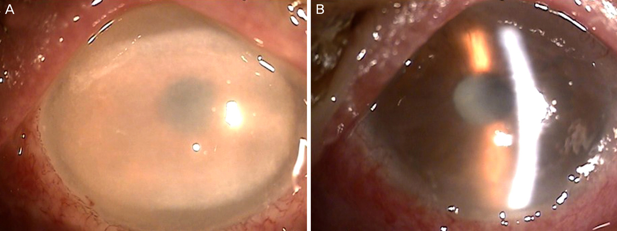
Figure 1. Slit-lamp photographs showing diffuse corneal edema and total epithelial defect with stromal infiltration at the cornea. (A) Right eye. (B) Left eye.
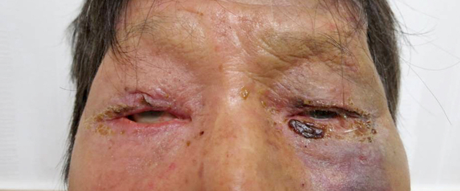
Figure 2. Clinical photograph of a 72-year-old male with both periocular redness and swelling with eschar change at the inferomedial area in his left lower eyelid.
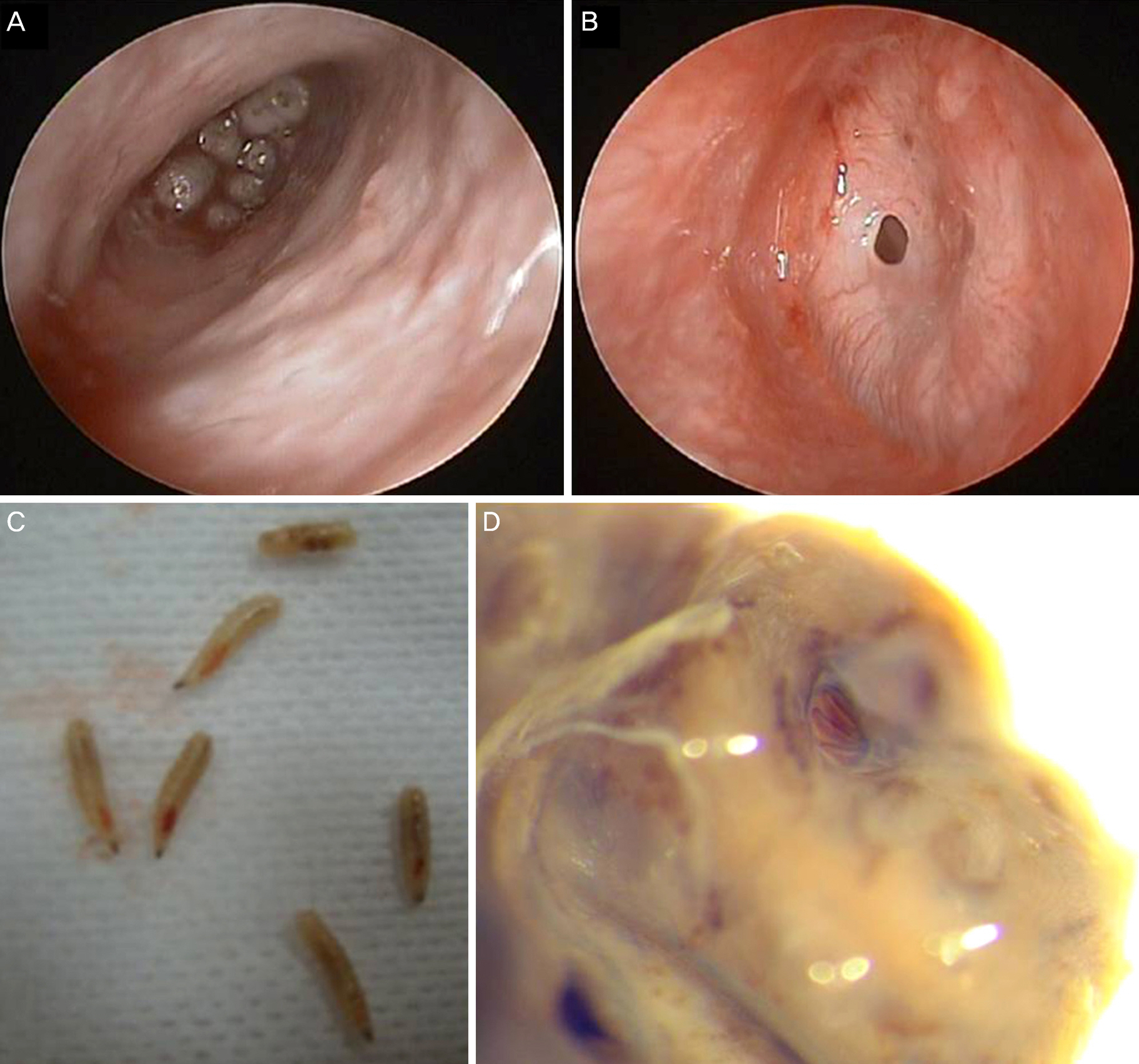
Figure 3. Otoscopic findings. (A) Numerous motile larvae with mucosal hyperemia and mucoid discharge are observed. (B) Ear drum perforation is observed after removal of larvae. (C) Six larvae were completely removed from the external acoustic meatus. (D) Microscopic findings of the removed larvae revealed L. sericata in their third stage of development. L. sericata = Lucilia sericata.
Reference
-
References
1. Keller AP Jr, Keller AP 3rd. Myiasis of the middle ear. Laryngoscope. 1970; 80:646–50.
Article2. Thompson JH, Knutson LV, Culp OS. Larva of Scenopinus sp. (diptera: scenopinidae) causing human urogenital myiasis? Mayo Clin Proc. 1970; 45:597–601.3. Misra N, Gogri P, Misra S, et al. Orbital myiasis caused by green bottle fly. Australas Med J. 2013; 6:504–6.
Article4. Holt GG, Adams TS, Sundet WD. Attraction and ovipositional response of screwworms, Cochliomyia hominivorax (Diptera: Calliphoridae), to stimulated bovine wounds. J Med Entomol. 1979; 16:248–53.5. Kim JS, Kim JW, Lee HJ, et al. Ophthalmomyiasis caused by a Phormia sp. (Diptera: Calliphoridae) larva in an enucleated patient. Korean J Parasitol. 2011; 49:173–5.6. Kalezić T, Stojković M, Vuković I, et al. Human external ophthalmomyiasis caused by Lucilia sericata Meigen (Diptera: Calliphoridae)-a green bottle fly. J Infect Dev Ctries. 2014; 8:925–8.7. Choi W, Kim GE, Park SH, et al. First report of external ophthalmomyiasis caused by Lucilia sericata Meigen in a healthy patient without predisposing risk factors. Parasitol Int. 2015; 64:281–3.
Article8. Navidpour SH. Myiasis in Khoozestan, “epidemiology, parasitology and control”. Khoozestan: Research Center of Natural Resources and Animal Husbandry. 1995; 35–51.9. Puthran N, Hegde V, Anupama B, Andrew S. Ivermectin treatment for massive orbital myiasis in an empty socket with concomitant scalp pediculosis. Indian J Ophthalmol. 2012; 60:225–7.
Article10. GS AM, SH, et al. External ophthalmomyiasis which was caused by sheep botfly (Oestrus ovis) larva: a report of 10 cases. J Clin Diagn Res. 2013; 7:539–42.
- Full Text Links
-
- Actions
-
Cited
- CITED
-
- Close
- Share
-
- Similar articles
-
- Nosocomial submandibular infections with dipterous fly larvae
- Traumatic Myiasis Caused by an Association of Sarcophaga tibialis (Diptera: Sarcophagidae) and Lucilia sericata (Diptera: Calliphoridae) in a Domestic Cat in Italy
- External Ophthalmomyiasis Presenting to an Emergency Department: Corneal Findings as a Sign of Oestrus ovis
- A Case of Oral Myiasis Caused by Lucilia sericata (Diptera: Calliphoridae) in Korea
- External Ophthalmomyiasis Caused by Oestrus ovis: A Rare Case Report from India
