Isolated primary schwannoma arising on the colon: report of two cases and review of the literature
- Affiliations
-
- 1Department of Surgery, Dong-A University College of Medicine, Busan, Korea. colonch@donga.ac.kr
- 2Department of Internal Medicine, Dong-A University College of Medicine, Busan, Korea.
- 3Department of Pathology, Dong-A University College of Medicine, Busan, Korea.
- KMID: 2212163
- DOI: http://doi.org/10.4174/jkss.2011.80.5.367
Abstract
- Primary schwannoma of the large intestine is an extremely rare neoplasm. Here, we report two cases of colonic schwannoma confirmed pathologically after laparoscopic resection. A 52-year-old female and a 59-year-old female were referred by their general practitioners to our coloproctologic clinic for further evaluation and management of colonic submucosal masses. Colonoscopies performed in our institution revealed round submucosal tumors with a smooth and intact mucosa in the mid-ascending and descending colon, respectively. Computed tomography (CT) scans showed an enhancing soft tissue mass measuring 2 x 2 cm in the right colon and well-defined soft tissue nodule measuring 1.5 x 1.7 cm in the proximal descending colon, respectively. We performed laparoscopic right hemicolectomy and segmental left colectomy under the preoperative impression of gastrointestinal stromal tumors. Two cases were both diagnosed to be benign schwannoma of the colon after immunohistochemical stains (S-100 (+), smooth muscle actin (-), CD117 (-), and CD34 (-)).
Keyword
MeSH Terms
Figure
-
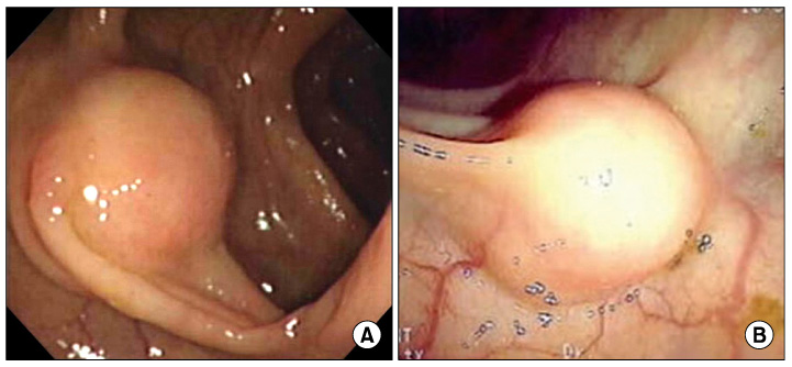
Fig. 1 Colonoscopic findings of both cases showing submucosal tumors measuring approximately 2 cm (A) and 1.5 cm (B) in diameter.

Fig. 2 Computed tomography scans. (A, B) Images of the first case show a well-defined homogeneously enhancingsubmucosal mass in the posterior wall of the cecum (arrow). (C, D) A well-defined soft tissue nodule can be seen in the proximal descending colon (arrow).
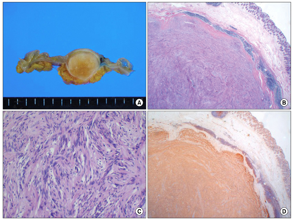
Fig. 3 Pathological examinations. (A) Ascending colon shows a well-circumscribed mass, measuring 1.8 × 1.6 cm. The mass is intramurally located and shows whitish fibrotic and firm cut surface without definitive hemorrhage or necrosis. The covering mucosa is intact. (B) The well-circumscribed intramural mass is surrounded by lymphocytic rim (H&E, ×10). (C) The mass is composed of whorled arranged spindle cells and the tumor cells are uniform and show few mitotic figures (H&E, ×100). (D) The tumor shows a diffuse cytoplasmicimmunoreactivity for S-100 protein (×10).
Reference
-
1. Daimaru Y, Kido H, Hashimoto H, Enjoji M. Benign schwannoma of the gastrointestinal tract: a clinicopathologic and immunohistochemical study. Hum Pathol. 1988. 19:257–264.2. Skopelitou AS, Mylonakis EP, Charchanti AV, Kappas AM. Cellular neurilemoma (schwannoma) of the descending colon mimicking carcinoma: report of a case. Dis Colon Rectum. 1998. 41:1193–1196.3. Hou YY, Tan YS, Xu JF, Wang XN, Lu SH, Ji Y, et al. Schwannoma of the gastrointestinal tract: a clinicopathological, immunohistochemical and ultrastructural study of 33 cases. Histopathology. 2006. 48:536–545.4. Miettinen M, Lasota J. Gastrointestinal stromal tumors--definition, clinical, histological, immunohistochemical, and molecular genetic features and differential diagnosis. Virchows Arch. 2001. 438:1–12.5. Miettinen M, Shekitka KM, Sobin LH. Schwannomas in the colon and rectum: a clinicopathologic and immunohistochemical study of 20 cases. Am J Surg Pathol. 2001. 25:846–855.6. Inagawa S, Hori M, Shimazaki J, Matsumoto S, Ishii H, Itabashi M, et al. Solitary schwannoma of the colon: report of two cases. Surg Today. 2001. 31:833–838.7. Levy AD, Quiles AM, Miettinen M, Sobin LH. Gastrointestinal schwannomas: CT features with clinicopathologic correlation. AJR Am J Roentgenol. 2005. 184:797–802.8. Mulchandani MH, Chattopadhyay D, Obafunwa JO, Joypaul VB. Gastrointestinal autonomic nerve tumours--report of a case and review of literature. World J Surg Oncol. 2005. 3:46.9. Lauwers GY, Erlandson RA, Casper ES, Brennan MF, Woodruff JM. Gastrointestinal autonomic nerve tumors. A clinicopathological, immunohistochemical, and ultrastructural study of 12 cases. Am J Surg Pathol. 1993. 17:887–897.10. Braumann C, Guenther N, Menenakos C, Junghans T. Schwannoma of the colon mimicking carcinoma: a case report and literature review. Int J Colorectal Dis. 2007. 22:1547–1548.
- Full Text Links
-
- Actions
-
Cited
- CITED
-
- Close
- Share
-
- Similar articles
-
- Isolated Colonic Schwannoma in the Ascending Colon: A Case Report and Literature Review of Schwannomas in the Large Intestine
- Melanotic Schwannoma in Cervical Spine: A Case Report
- Surgical treatment of multiple plexiform schwannomas arising from the superficial radial nerve: a case report
- A Case of Schwannoma of the Tonsil
- A Case of Schwannoma Arising in Pterygopalatine Fossa
