J Korean Ophthalmol Soc.
2008 Aug;49(8):1317-1322. 10.3341/jkos.2008.49.8.1317.
Analysis of Refractive Error and Corneal Asphericity in Elementary School Students in Ilsan City
- Affiliations
-
- 1Department of Ophthalmology and Visual Science, College of Medicine, The Catholic University of Korea, Seoul, Korea. yclee@cmcnu.or.kr
- 2Graduate School of Public Health, The Eulji University of Korea, Daejeon, Korea.
- KMID: 2211738
- DOI: http://doi.org/10.3341/jkos.2008.49.8.1317
Abstract
- PURPOSE
To determine the relationships among refractive error, corneal asphericity, and axial length in elementary school students.
METHODS
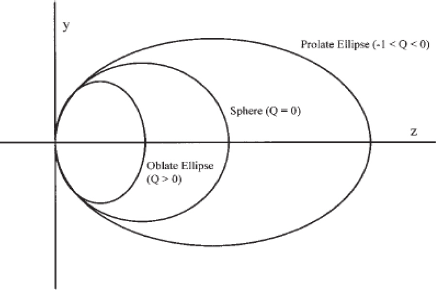
One hundred eleven eyes from 56 subjects were included in this study. All subjects underwent cycloplegic refraction corrected to the spherical equivalent. Axial length was measured, and corneal topography was performed. Corneal asphericity was assessed using eccentricity (e) calculated according to the formula Q=-e2. The relationship among spherical equivalent, asphericity, and axial length was determined using a linear regression model.
RESULTS
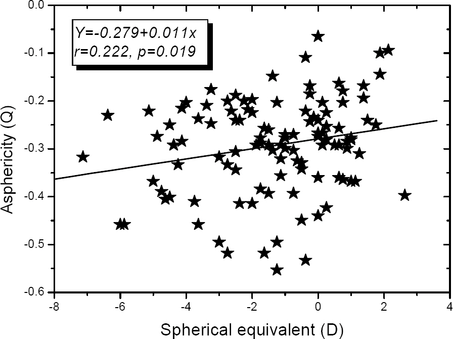
Subjects were between 8 and 12 years of age (mean, 9.99+/-1.33). The average spherical equivalent was -1.38+/-2.08D (-7.13~2.63D), the average axial length was 23.84+/-1.17 mm (20.10~26.37 mm), and the average corneal asphericity was -0.29+/-0.10 (-0.55~-0.07). An increase in myopia was positively correlated with an increase in axial length (p<0.0001). The degree of myopia was negatively associated with corneal asphericity (p=0.019). An increase in axial length was related to an increase of negativity in asphericity (p=0.012).
CONCLUSIONS
An increase in myopia was correlated with an increase in axial length. As the degree of myopia and axial length increased, corneal asphericity became more prolate. A longitudinal study with more subjects is required to validate these results.
Keyword
Figure
Reference
-
References
1. Mandell RB. Everett Kinsey Lecture. The enigma of the corneal contour. CLAO J. 1992; 18:267–73.2. Mandell RB, St Helen R. Mathematical model of the corneal contour. Br J Physiol Opt. 1971; 26:183–197.3. Davis WR, Raasch TW, Mitchell GL. . Corneal asphericity and apical curvature in children: A cross-sectional and longitudinal evaluation. Invest Ophthalmol Vis Sci. 2005; 46:1899–906.
Article4. Kiely PM, Smith G, Carney LG. The mean shape of the human cornea. Optica Acta. 1982; 29:1027–40.
Article5. Liou HL, Brennan NA. Anatomically accurate, finite model eye for optical modeling. J Opt Soc Am A Opt Image Sci Vis. 1997; 14:1684–95.
Article6. Horner DG, Soni PS, Vyas N, Himebaugh NL. Longitudinal changes in corneal asphericity in myopia. Optom Vis Sci. 2000; 77:198–203.
Article7. Lindsay R, Smith G, Atchison D. Descriptors of corneal shape. Optom Vis Sci. 1998; 75:156–8.
Article8. Kim JC, Koo BS. A study of prevailing features and causes of myopia and visual impairment in urban school children. J Korean Ophthalmol Soc. 1988; 29:165–81.9. Koo BS, Kim JC, Chung HS. A study of the ocular findings according to subdivided myopia. J Korean Ophthalmol Soc. 1987; 28:133–8.10. Millodot M, Sivak J. Contribution of the cornea and lens to the spherical aberration of the eye. Vision Res. 1979; 19:685–7.
Article11. Eghbali F, Yeung KK, Maloney RK. Topographic determination of corneal asphericity and its lack of effect on the refractive outcome of radial keratotomy. Am J Ophthalmol. 1995; 119:275–80.
Article12. Lam AK, Douthwaite WA. Application of a modified keratometer in the study of corneal topography on Chinese subjects. Ophthalmic Physiol Opt. 1996; 16:130–4.
Article13. Townsley MG. New knowledge of the corneal contour. Contacto. 1970; 14:38–43.14. Kiely PM, Smith G, Carney LG. Meridional variations of corneal shape. Am J Optom Physiol Opt. 1984; 61:619–26.
Article15. Carney LG, Mainstone JC, Henderson BA. Corneal topography and myopia, A cross-sectional study. Invest Ophthalmol Vis Sci. 1997; 38:311–20.16. Carney LG. The basis for corneal shape change during contact lens wear. Am J Optom Physiol Opt. 1975; 52:445–54.
Article17. Dave T, Ruston D. Current trends in modern orthokeratology. Ophthalmic Physicol Opt. 1998; 18:224–33.
Article18. Swarbrick HA, Wong G, O'Leary DJ. Corneal response to orthokeratology. Optom Vis Sci. 1998; 75:791–9.19. Kang SY, Kim BK, Byun YJ. Sustainability of orthokeratology as demonstrated by corneal topography. Korean J Ophthalmol. 2007; 21:74–8.
Article20. Chang JW, Choi TH, Lee HB. The efficacy and safety of reverse geometry lenses. J Korean Ophthalmol Soc. 2004; 45:908–12.21. Nichols JJ, Marsich MM, Nguyen M. . Overnight orthokeratology. Optom Vis Sci. 2000; 77:252–9.
Article
- Full Text Links
- Actions
-
Cited
- CITED
-
- Close
- Share
- Similar articles
-
- Central Corneal Thickness Measured by Ultrasonic Pachymeter in Normal Koreans
- A Study of the Ocular Findings According to Subdivided Myepia
- A survey of the Refractive State of Elementary School Children in Rural Area
- The Relationship Between Asphericity and Visual Acuity After Wearing Reverse-Geometry Lens
- The Effect of Strabismus Surgery on Refractive Error Measured with Corneal Topography


