J Korean Ophthalmol Soc.
2008 Aug;49(8):1303-1308. 10.3341/jkos.2008.49.8.1303.
Comparison between Heidelberg Retina Tomograph 2 and Heidelberg Retina Tomograph 3 for Glaucoma Detection
- Affiliations
-
- 1Department of Ophthalmology and Visual Science, College of Medicine, The Catholic University of Korea, Seoul, Korea. ckpark@catholic.ac.kr
- KMID: 2211736
- DOI: http://doi.org/10.3341/jkos.2008.49.8.1303
Abstract
- PURPOSE
To compare the ability of Heidelberg Retina Tomograph (HRT) 2 and the new version, HRT3, to discriminate between glaucoma and non-glaucoma eyes and to evaluate the most useful parameters for glaucoma detection.
METHODS
Fifty-two healthy eyes, 62 glaucoma-suspect eyes, and 172 glaucoma eyes (a total of 286 eyes) were studied retrospectively. The discrimination capabilities of healthy and glaucomatous eyes were compared using areas under the receiver operating characteristics (AROCs) curves. Agreement between classifications was defined by kappa analysis.
RESULTS
The stereometric parameters that best discriminated between glaucoma and non-glaucoma eyes were cup shape measure at HRT2 (areas under the ROCs curves (AUC) 0.784) and global probability score (GPS) at HRT3 (AUC 0.718). The stereometric parameters that best discriminated between normal and non-normal eyes were cup shape measure at HRT2 (AUC 0.785) and GPS at HRT3 (AUC 0.696). The sensitivity was 95% at HRT3 and 91% at HRT2, and specificity was 61% at HRT2 and 59% at HRT3.
CONCLUSIONS
The glaucoma-discriminating ability of the HRT3 software is similar to that of HRT2; however, HRT3 provided results without the need for subjective operator input.
MeSH Terms
Figure
-

Figure 1. Comparision of glaucoma and non-glaucoma eyes with HRT (Heidelberg Retina tomography) 2.
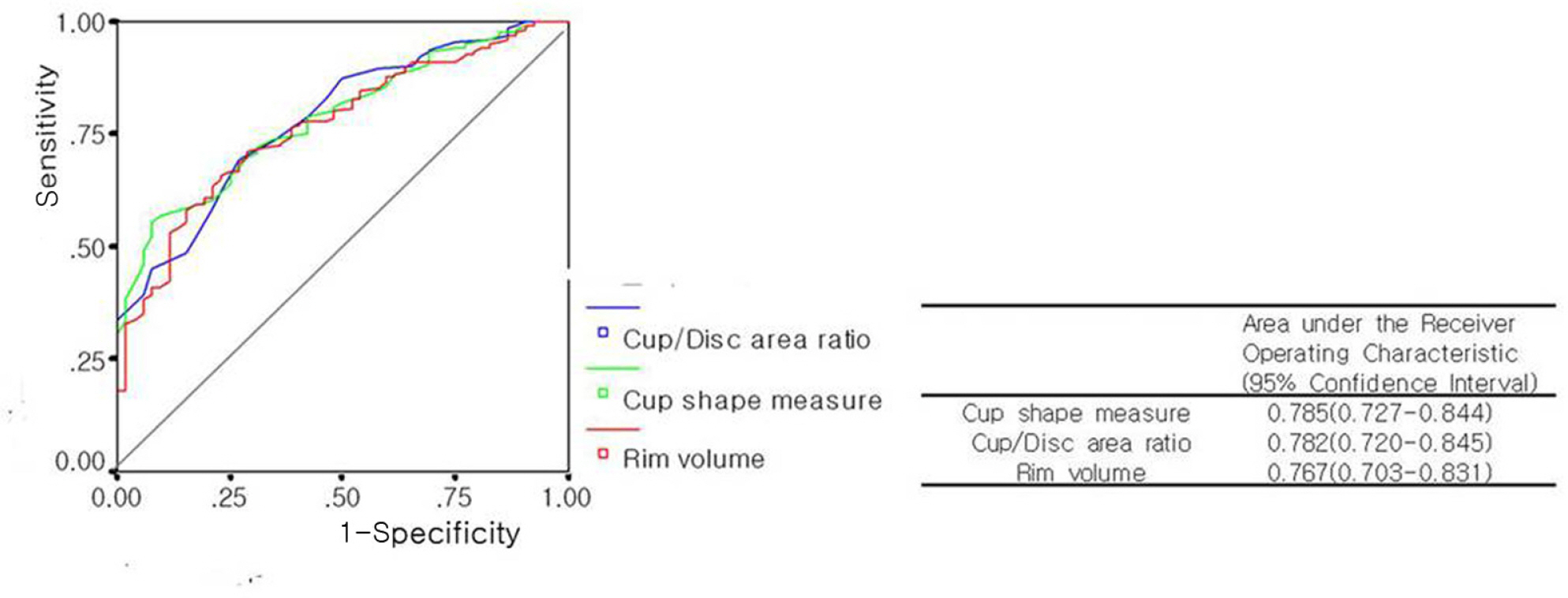
Figure 2. Comparision of glaucoma and non-glaucoma eyes with HRT (Heidelberg Retina Tomograph) 3.* GPS=Global probability score.

Figure 3. Comparision of normal and non-normal eyes with HRT (Heidelberg Retina Tomograph) 2.
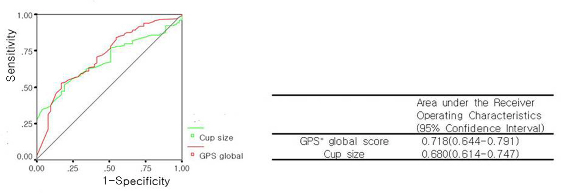
Figure 4. Comparision of normal and non-normal eyes with HRT (Heidelberg Retina Tomograph) 3.* GPS=Global probability score
Reference
-
References
1. Quigley HA. Number of people with glaucoma worldwide. Br J Ophthalmol. 1996; 80:389–93.
Article2. Kruse FE, Burk RO, Volcker HE. . Reproducibility of topographic measurements of the optic nerve head with laser tomographic scanning. Ophthalmology. 1989; 96:1320–4.
Article3. Dreher AW, Tse PC, Weinreb RN. Reproducibility of topographic measurements of the normal and glaucomatous optic nerve head with the laser tomographic scanner. Am J Ophthalmol. 1991; 111:221–9.
Article4. Rohrschneider K, Burk RO, Volcker HE. Reproducibility of topometric data acquisition in normal and glaucomatous optic nerve heads with the laser tomographic scanner. Graefes Arch Clin Exp Ophthalmol. 1993; 231:457–64.
Article5. Miqlior S, Casula M, Guareschi M. . Clinical ability of Heidelberg retinal tomograph examination to detect glauco matous visual field changes. Ophthalmology. 2001; 108:1621–7.6. Uchida H, Brigatti L, Caprioli J. Detection of structural damage from glaucoma with confocal laser image analysis. Invest Ophthalmol Vis Sci. 1996; 37:2393–401.7. Hatch WV, Flanagan JG, Etchells EE. . Laser scanning tomogrphy of the optic nerve head in ocular hypertension and glaucoma. Br J Ophthalmol. 1997; 104:545–8.8. Miglior S, Albe E, Guareschi M. . Intraobserver and interobserver reproducibility in the evaulation of optic disc steremetric parameters by Heidelberg Retina Tomograph. Ophthalmology. 2002; 109:1072–7.9. Garway-Heath DF, Poinoosawmy D, Wollstein G. . Interobserver and intraobserver variation in the analysis of optic disc images : comparision of the Heidelberg retina tomograph and computer assisted planimetry. Br J Ophthalmol. 1999; 83:664–9.10. Iester M, Mikelberg FS, Courtright P. . Interobserver variability of optic disc variables measured by confocal scanning laser tomography. Am J Ophthalmol. 2001; 132:57–62.11. Swindale NV, Stjepanovic G, Chin A, Mikelberg FS.Automated analysis of normal and glaucomatous optic nerve head topography images. Invest Ophthalmol Vis Sci. 2000; 41:1730–42.12. Park YJ, Park CK, Moon JI, Baek NH. The Diagnostic Precision of Glaucoma Classification with New HRT Discriminant Formula in Koreans. J Korean Ophthalmol Soc. 1999; 40:175–81.13. Sommer A, Pollack I, Maumence AE. Optic disc parameters and onset of glaucomatous field loss. Arch Ophthalmol. 1979; 97:1444–8.
Article14. Quigley HA, Addicks EM, Gree WR. Optic nerve damage in human glaucoma. Quantitative correlation of nerve fiber loss and visual field defect in glaucoma, ischemic neuropathy, papilledema, and toxic neuropathy. Arch Ophthalmol. 1982; 100:135–46.15. Pederson JE, Anderson DR. The mode of progressive disc cupping in ocular hypertension and glaucoma. Arch Ophthalmol. 1980; 98:490–5.
Article16. Caprioli J, Miller JM, Sears M. Quantitative evaluation of the optic nerve head in patients with unilateral visual field loss from primary open angle glaucoma. Ophthalmology. 1987; 94:1484–7.
- Full Text Links
-
- Actions
-
Cited
- CITED
-
- Close
- Share
-
- Similar articles
-
- Reproducibility of Optic Nerve Head Topographic Measurements with the Heidelberg Retina Tomograph
- Comparison of Optic Disc Measurements between Computer-aided Morphometry and Heidelberg Retina Tomograph
- Early Detection of Glaucoma with Retinal Nerve Fiber Layer Photograph
- Reversibility of Optic Disc Cupping after Trabeculectomy in Adult Glaucoma Patients Measured by Heidelberg Retina Tomograph
- The Effect of Centrally Aligned Image Acquisition on the Reproducibility of Optic Nerve Head Topographic Parameters obtained with Heidelberg Retina Tomograph
