J Korean Ophthalmol Soc.
2008 Jan;49(1):119-124. 10.3341/jkos.2008.49.1.119.
The Effect of Myopic Optical Defocus on the Humphrey Matrix 30-2 Test
- Affiliations
-
- 1Department of Ophthalmology, Samsung Medical Center, Sungkyunkwan University School of Medicine, Seoul, Korea. cwkee@smc.samsung.co.kr
- KMID: 2211191
- DOI: http://doi.org/10.3341/jkos.2008.49.1.119
Abstract
-
PURPOSE: To investigate the effect of myopic optical defocus on Humphrey Matrix 30-2 test.
METHODS
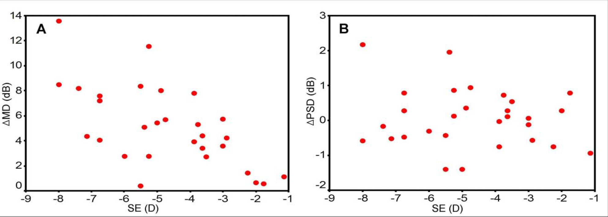
Twenty-nine myopic eyes of 29 patients underwent 2 consecutive tests with the Humphrey Matrix 30-2 threshold program. The first and second tests were performed with and without correction for myopia. Differences between the mean deviation (delta MD) and the pattern standard deviation (delta PSD) were calculated, and a correlation between the spherical equivalent (SE) and delta MD and delta PSD was investigated. The influence of optical defocus according to the visual field (central and peripheral) and the severity of glaucomatous visual field damage (area with total deviation plot of 'P<0.1%' or 'P<0.5%' and the other area) were also evaluated.
RESULTS
The correlation between SE and delta MD was significant (p=0.02, r=0.62). However, the correlation between SE and delta PSD was not significant (p=0.81, r=0.15). The differences in the mean total deviation printout value of the central and peripheral visual field groups between the two tests were 5.85+/-4.26dB and 5.66+/-3.56dB, respectively (P=0.86). The differences in the mean total deviation printout value of severely damaged and less damaged areas between the two tests were 3.16+/-3.39dB and 6.62+/-4.62dB, respectively (P<0.01).
CONCLUSIONS
Myopic optical defocus has a significant effect on the results of the Humphrey Matrix 30-2 test. There is no difference in the effect of myopia between the central and peripheral visual fields, and the effect is decreased in severely damaged visual fields.
MeSH Terms
Figure
Cited by 1 articles
-
The Effect of One Myopic Eye on Visual Field Testing
Yong Kyun Shin, Myung Won Lee, Kyoung Jin Cho, Moo Hwan Chang
J Korean Ophthalmol Soc. 2014;55(9):1355-1360. doi: 10.3341/jkos.2014.55.9.1355.
Reference
-
References
1. Quigley HA, Dunkelberger GR, Green WR. Retinal ganglion cell atrophy correlated with automated perimetry in human eyes with glaucoma. Am J Ophthalmol. 1989; 107:453–64.
Article2. Kerrigan-Baumrind LA, Quigley HA, Pease ME, et al. Number of ganglion cells in glaucoma eyes compared with threshold visual field tests in the same persons. Invest Ophthalmol Vis Sci. 2000; 41:741–8.3. Johnson CA, Adams AJ, Casson EJ, et al. Blue-on-yellow perimetry can predict the development of glaucomatous visual field loss. Arch Ophthalmol. 1993; 111:645–50.
Article4. Johnson ca, Adams AJ, Casson EJ, Brandt JD. Progression of early glaucomatous visual field loss as detected by blue-on-yellow and standard white-on-white automated perimetry. Arch Ophthalmol. 1993; 111:651–6.
Article5. Wall M, Ketoff KM. Random dot motion perimetry in patients with glaucoma and in normal subjects. Am J Ophthalmol. 1995; 120:587–96.
Article6. Bosworth CF, Sample SA, Gupta N, et al. Motion automated perimetry identifies early glaucomatous field defects. Arch Ophthalmol. 1998; 116:1153–8.
Article7. Glovinsky Y, Quigley HA, Pease ME. Foveal ganglion cell loss in size dependent on experimental glaucoma. Invest Ophthalmol Vis Sci. 1991; 32:484–91.8. Quigley HA, Dunkelberger GR, Green WR. Chronic human glaucoma causing selectively greater loss of large optic nerve fibers. Ophthalmology. 1988; 95:357–63.
Article9. Kelly DH. Frequency doubling in visual responses. J Opt Soc Am. 1966; 56:1628–33.
Article10. Johnson CA, Samuels SJ. Screening for glaucomatous visual field loss with frequency-doubling perimetry. Invest Ophthalmol Vis Sci. 1997; 38:413–25.11. Quigley HA. Identification of glaucoma-related visual field abnormality with the screening protocol of frequency doubling technology. Am J Ophthalmol. 1998; 125:819–29.12. Brusini P, Salvetat ML, Zeppieri M, Parisi L. Frequency doubling technology perimetry with the Humphrey Matrix 30-2 test. J Glaucoma. 2006; 15:77–83.
Article13. Ko BS, Kim CY, Hong YJ. Correlation between optic disc tomography and frequency doubling technology in glaucoma suspect. J Korean Ophthalmol Soc. 2003; 44:2040–6.14. Anderson AJ, Johnson CA. Frequency-doubling technology perimetry and optical defocus. Invest Ophthalmol Vis Sci. 2003; 44:4147–52.
Article15. Artes PH, Nicolela MT, McCormick TA, et al. Effects of blur and repeated testing on sensitivity estimates with frequency doubling perimetry. Invest Ophthalmol Vis Sci. 2003; 44:646–52.
Article16. Ito A, Kawabata H, Fujimoto N, Adachi-Usami E. Effect of myopia on frequency-doubling perimetry. Invest Ophthalmol Vis Sci. 2001; 42:1107–10.17. Heeg GP, Stoutenbeek R, Jansonius NM. Strategies for improving the diagnostic specificity of the frequency doubling perimeter. Acta Ophthalmol Scand. 2005; 83:53–6.
Article18. Heeg GP, Ponsioen TL, Jansonius NM. Learning effect, normal range, and test-retest variability of Frequency Doubling Perimetry as a function of age, perimetric experience, and the presence or absence of glaucoma. Ophthalmic Physiol Opt. 2003; 23:535–40.
Article19. Joson PJ, Kamantigue ME, Chen PP. Learning effects among perimetric novices in frequency doubling technology perimetry. Ophthalmology. 2002; 109:757–60.
Article20. Iester M, Capris P, Pandolfo A, et al. Learning effect, short-term fluctuation, and long-term fluctuation in frequency doubling technique. Am J Ophthalmol. 2000; 130:160–4.
Article21. Brush MB, Chen PP. Test-retest variability in glaucoma patients tested with C-20-1 screening-mode frequency doubling technology perimetry. J Glaucoma. 2004; 13:273–7.
Article22. Adams CW, Bullimore MA, Wall M, et al. Normal aging effects for frequency doubling technology perimetry. Optom Vis Sci. 1999; 76:582–7.
Article23. Legge GE, Mullen KT, Woo GC, Campbell FW. Tolerance to visual defocus. J Opt Soc Am A. 1987; 4:851–63.
Article24. Brusini P, Salvetat ML, Zeppieri M, Parisi L. Frequency doubling technology perimetry with the Humphrey Matrix 30-2 test. J Glaucoma. 2006; 15:77–83.
Article25. Kogure S, Toda Y, Crabb D, et al. Agreement between frequency doubling perimetry and static perimetry in eyes with high tension glaucoma and normal tension glaucoma. Br J Ophthalmol. 2003; 87:604–8.
Article26. Anderson AJ, Johnson CA, Fingeret M, et al. Characteristics of the normative database for the Humphrey matrix perimeter. Invest Ophthalmol Vis Sci. 2005; 46:1540–8.
Article27. Haymes SA, Hutchison DM, McCormick TA, et al. Glaucomatous visual field progression with frequency-doubling technology and standard automated perimetry in a longitudinal prospective study. Invest Ophthalmol Vis Sci. 2005; 46:547–54.
Article
- Full Text Links
- Actions
-
Cited
- CITED
-
- Close
- Share
- Similar articles
-
- The Effect of One Myopic Eye on Visual Field Testing
- The Comparison of The Matrix Perimetry and Humphrey Standard Perimetry in Various Patients Group
- Effect of Dual-focus Contact Lenses on Myopic Progression
- Peripheral Defocus and Myopia Management: A Mini-Review
- Measuring Defocus Curves of Monofocal, Multifocal and Extended Depth-of-focus Intraocular Lenses Using Optical Bench Test


