Optimal Standing Radiographic Positioning in Patients with Sagittal Imbalance
- Affiliations
-
- 1Department of Orthopaedic Surgery, Eulji University School of Medicine, Daejeon, Korea. hjkim@eulji.ac.kr
- KMID: 2209583
- DOI: http://doi.org/10.4184/jkss.2010.17.4.198
Abstract
- STUDY DESIGN: This is a review of the literature about radiographic positioning for patients with sagittal imbalance.
OBJECTIVES
We wanted to verify the optimal radiographic positioning for patients with sagittal imbalance. SUMMARY OF LITERATURE REVIEW: The standing lateral whole spine radiograph for identifying the sagittal alignment has a different value for the SVA according to the radiographic positioning.
MATERIALS AND METHODS
This is a review of the literature.
RESULTS
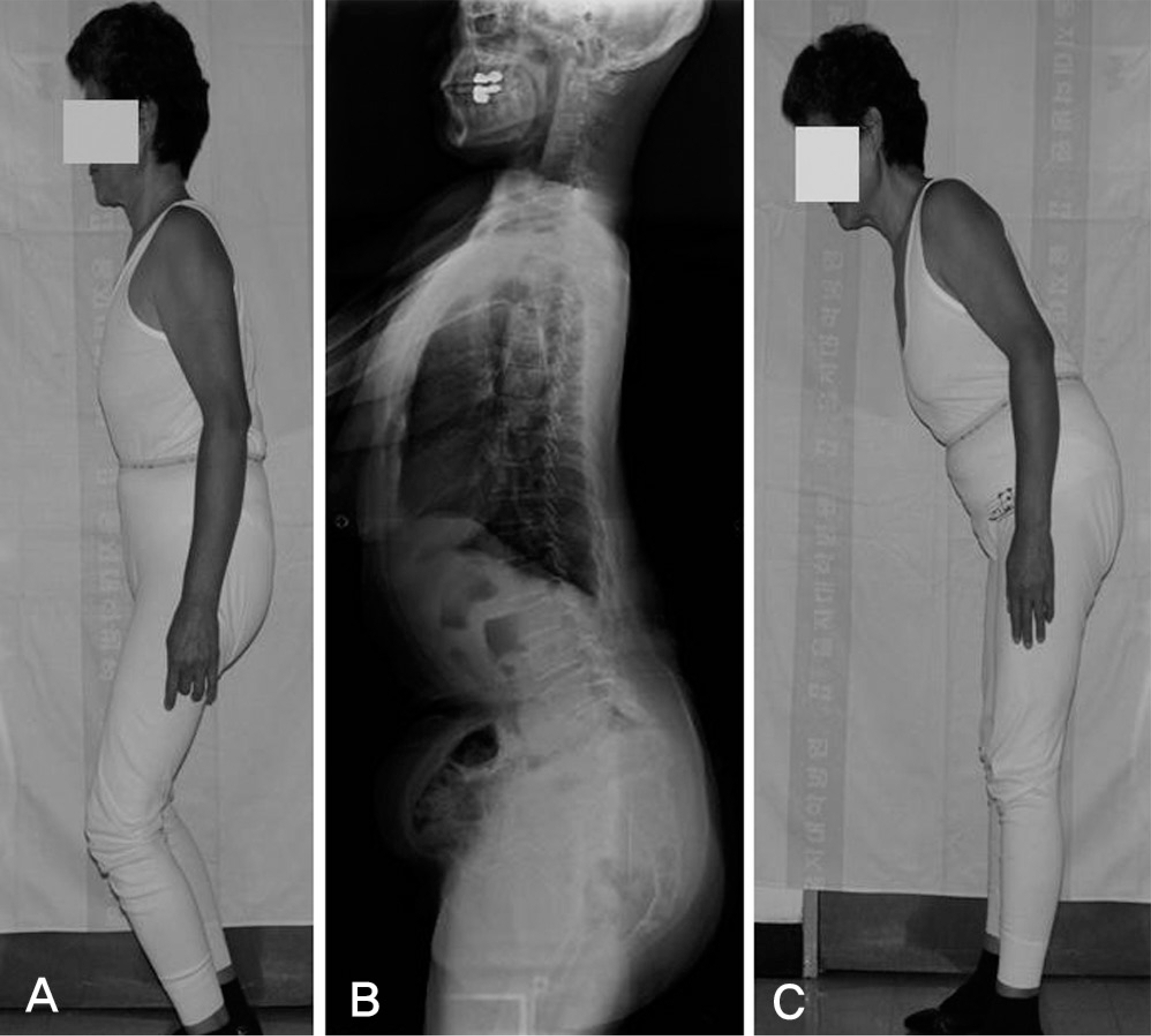
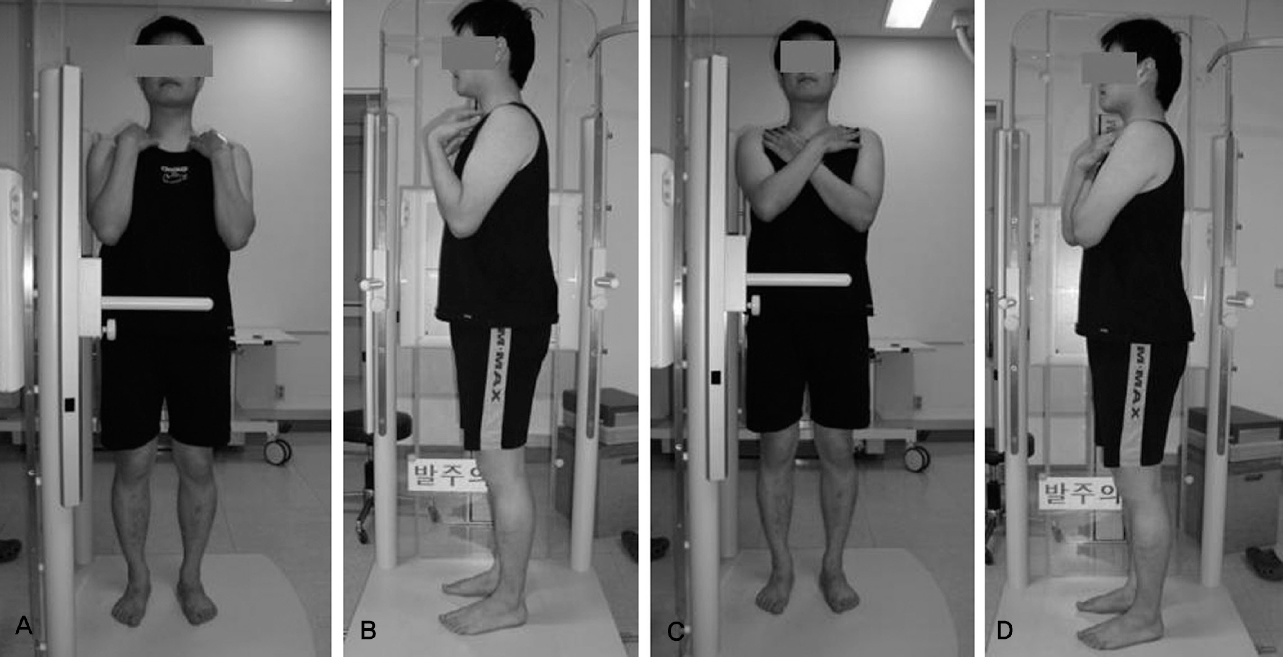
The fists-on-the clavicle position or the cross-arm position not only represents a functional standing position, but it also causes a less negative shift of the SVA in patients with sagittal imbalance. Both the extended hip and knee positions are necessary to exclude a compensation mechanism of the lower extremity.
CONCLUSIONS
The optimal radiographic positioning is essential to examine the degrees of sagittal imbalance.
Keyword
Figure
Cited by 3 articles
-
Results of Corrective Osteotomy and Treatment Strategy for Ankylosing Spondylitis with Kyphotic Deformity
Ki-Tack Kim, Dae-Hyun Park, Sang-Hun Lee, Jung-Hee Lee
Clin Orthop Surg. 2015;7(3):330-336. doi: 10.4055/cios.2015.7.3.330.Changes of Spinopelvic Parameter using Iliac Screw In Surgical Correction of Sagittal Imbalance Patients
Whoan Jeang Kim, Yong Joo Chi, Dae Geon Song, Kyung Hoon Park, Kun Young Park, Hwan Il Sung, Je Yun Koo, Won Cho Kwon, Won Sik Choy
J Korean Soc Spine Surg. 2014;21(2):63-69. doi: 10.4184/jkss.2014.21.2.63.Natural History of Lumbar Degenerative Kyphosis with Conservative Treatment
Whoan Jeang Kim, Shann Haw Chang, Gyu Sang Lee, Yong Ho Kim, Kun Young Park, Kyung Hoon Park, Won Sik Choy
J Korean Soc Spine Surg. 2017;24(1):24-31. doi: 10.4184/jkss.2017.24.1.24.
Reference
-
1.Duval-Beaupè re G., Schimdt C., Cosson P. A barycentremetric study of the sagittal shape of spine and pelvis: the conditions required for an economic standing position. Ann Biomed Eng. 1992. 20:451–62.2.Jackson RP., McManus AC. Radiographic analysis of sagittal plane alignment and balance in standing volunteers and patients with low back pain matched for age, sex, and size. A prospective controlled clinical study. Spine. 1994. 19:1611–8.3.Mannion AF., Taimela S., Mü ntener M., Dvorak J. Active therapy for chronic low back pain part 1. Effects on back muscle activation, fatigability, and strength. Spine. 2001. 26:897–908.4.Takahashi T., Ishida K., Hirose D, et al. Trunk deformity is associated with a reduction in outdoor activities of daily living and life satisfaction in community-dwelling older people. Osteoporos Int. 2005. 16:273–9.
Article5.Lee CS., Kim YT., Kim E. Clinical study of lumbar degenerative kyphosis. J Korean Soc Spine Surg. 1997. 4:27–35.6.Gelb DE., Lenke LG., Bridwell KH., Blanke K., McEnery KW. An analysis of sagittal spinal alignment in 100 asymptomatic middle and older aged volunteers. Spine. 1995. 20:1351–8.
Article7.Kim WJ., Kang JW., Yeom JS, et al. A comparative analysis of sagittal spinal balance in 100 asymptomatic young and older aged volunteers. J Korean Soc Spine Surg. 2003. 10:327–34.
Article8.Lee CS., Oh WH., Chung SS., Lee SG., Lee JY. Analysis of the sagittal alignment of normal spines. J Korean Orthop Assoc. 1999. 34:949–54.
Article9.Lafage V., Schwab F., Skalli W, et al. Standing balance and sagittal plane spinal deformity: analysis of spinopelvic and gravity line parameters. Spine. 2008. 33:1572–8.10.Hammerberg EM., Wood KB. Sagittal profile of the elderly. J Spinal Disord Tech. 2003. 16:44–50.
Article11.Stagnara P., De Mauroy JC., Dran G, et al. Reciprocal angulation of vertebral bodies in a sagittal plane: approach to references in the evaluation of kyphosis and lordosis. Spine. 1982. 7:335–42.12.Kim MS., Chung SW., Hwang CJ., Lee CK., Chang BS. A radiographic analysis of sagittal spinal alignment for the standardization of standing lateral position. J Korean Orthop Assoc. 2005. 40:861–7.
Article13.Vendatum R., Lenke LG., Keeney JA., Bridwell KH. Comparison of standing sagittal spinal alignment in asymptomatic adolescents and adults. Spine. 1998. 23:211–5.14.Peterson MD., Jackson RP., McManus AC. Standing sagittal spinal balance, alignments and lumbopelvic relationships: Part I. A study of adult volunteers. Presented at the annual meeting of the Scoliosis Research Society, Asheville, North Carolina, September. 1995. 13–7.15.Legaye J., Duval-Beaupere G., Hecquet J., Marty C. Pelvic incidence: a fundamental pelvic parameter for three-dimensional regulation of spinal sagittal curves. Eur Spine J. 1998. 7:99–103.
Article16.Labelle H., Roussouly P., Berthonnaud E, et al. Spondylolisthesis, pelvic incidence, and spinopelvic balance: a correlation study. Spine. 2004. 29:2049–54.17.Mendoza-Lattes S., Ries Z., Gao Y., Weinstein SL. Natural history of spinopelvic alignment differs from symptomatic deformity of the spine. Spine. 2010. 35:E792–8.
Article18.Jackson RP., Kanemura T., Kawakami N., Hales C. Lumbopelvic lordosis and pelvic balance on repeated standing lateral radiographs of adult volunteers and untreated patients with constant low back pain. Spine. 2000. 25:575–86.
Article19.Marks MC., Stanford CF., Mahar AT., Newton PO. Standing lateral radiographic positioning does not represent customary standing balance. Spine. 2003. 28:1176–82.
Article20.Aota Y., Saito T., Uesugi M., Ishida K., Shinoda K., Mizuma K. Does the fists-on-clavicles position represent a functional standing position? Spine. 2009. 34:808–12.
Article21.Faro FD., Marks MC., Pawelek J., Newton PO. Evaluation of a functional position for lateral radiograph acquisition in adolescent idiopathic scoliosis. Spine. 2004. 29:2284–9.
Article22.Kim WJ., Kang JW., Koo JY, et al. Changes of sagittal alignment according to position in patients with degenerative kyphotic deformity. The 54th Annual Fall Congress of the Korean Orthopaedic Association. Oct, 2010. (Oral presentation).23.Horton WC., Brown CW., Bridwell KH., Glassman SD., Suk SI., Cha CW. Is there an optimal patient stance for obtaining a lateral 36” radiograph? a critical comparison of three techniques. Spine. 2005. 30:427–33.24.Suzuki H., Endo K., Mizuochi J., Kobayashi H., Tanaka H., Yamamoto K. Clasped position for measurement of sagittal spinal alignment. Eur Spine J. 2010. 19:782–6.
Article25.O'Brien MF., Kuklo TR., Blanke KM., Lenke LG. Radiographic Measurement Manual. Spinal Deformity Study Group(SDSG). Medtronic Sofamor Danek. 2004.26.Jackson RP., Hales C. Congruent spinopelvic alignment on standing lateral radiographs of adult volunteers. Spine. 2000. 25:2808–15.
Article27.Kim WJ., Kang JW., Kim HY, et al. Change of pelvic tilt before and after gait in patients with lumbar degenerative kyphosis. J Korean Soc Spine Surg. 2009. 16:95–103.
Article28.Schwab F., Lafage V., Boyce R., Skalli W., Farcy JP. Gravity line analysis in adult volunteers. age-related correlation with spinal parameters, pelvic parameters, and foot position. Spine. 2006. 31:E959–67.
- Full Text Links
- Actions
-
Cited
- CITED
-
- Close
- Share
- Similar articles
-
- Comparison of Surgical Burden, Radiographic and Clinical Outcomes According to the Severity of Baseline Sagittal Imbalance in Adult Spinal Deformity Patients
- Sagittal Imbalance
- Necessity of Whole Spine Standing Lateral Radiograph in Adults over 50 Years Old Who Have Degenerative Lumbar Disease: Comparison with Supine Lumbar Lateral Radiograph
- Correlation of Sagittal Imbalance and Recollapse after Percutaneous Vertebroplasty for Thoracolumbar Osteoporotic Vertebral Compression Fracture: A Multivariate Study of Risk Factors
- Sagittal Imbalance


