Synovial Chondrosarcoma in the Hand and Wrist: A Case Report
- Affiliations
-
- 1Department of Radiology, St. Vincent's Hospital, College of Medicine, The Catholic University of Korea, Korea. jhb@dreamwiz.com
- 2Department of Pathology, St. Vincent's Hospital, College of Medicine, The Catholic University of Korea, Korea.
- 3Department of Orthopedic Surgery, St. Vincent's Hospital, College of Medicine, The Catholic University of Korea, Korea.
- KMID: 2208998
- DOI: http://doi.org/10.3348/jksr.2010.62.1.73
Abstract
- Synovial chondrosarcoma is extremely rare and arises de novo or from malignant transformation of synovial chondromatosis. It commonly involves large joints, such as the knee or hip. Here, we present an unusual case of synovial chondrosarcoma from synovial chondromatosis in the hand and wrist, clearly demonstrating the characteristic findings on plain radiograph and MR imaging.
MeSH Terms
Figure
-
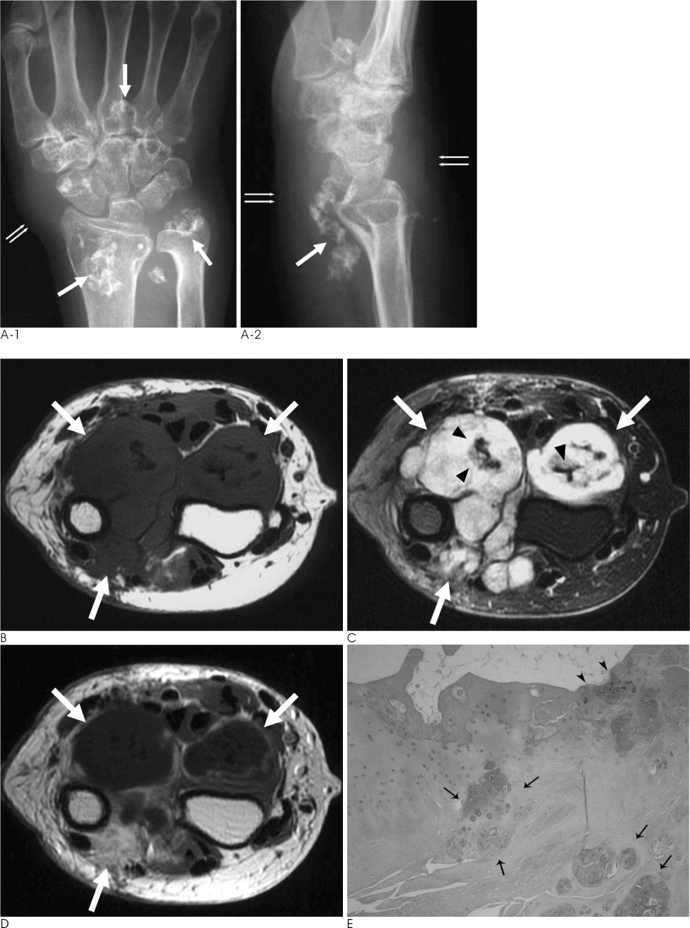
Fig. 1 Synovial chondrosarcoma in an 82-year-old woman. A. Plain radiograph shows multiple, popcorn-like with ring- and arc-shaped calcifications (thick arrows) in the wrist and proximal hand. Marked soft tissue swelling (thin double arrows) is seen in this area. B. T1-weighted axial image shows a large lobulated, hypointense mass (white arrows) from the distal metaphysis of the radius and ulna to the metaphysis of the metacarpal bones. C. This mass is hyperintense on fat suppressed T2-weighted axial image (white arrows). The medullary canal is intact. Note multiple dark signal intensity nodules within the mass (black arrowheads), suggesting chondroid calcifications on plain radiograph. D. Contrast enhanced T1-weighted image shows nodular and septal enhancement in this mass (white arrows). E. Photomicrograph (Hematoxylin-eosin stain: original magnification, ×100) depicts a lobular cartilage mass with infiltration into surrounding soft tissue and abutment to the adjacent bone cortex. Note the malignant tumor cells nests (black thin arrows). This malignant tumor cell nest focally invades into the cortex of adjacent bone (black arrowheads).
Reference
-
1. Bertoni F, Unni KK, Beabout JW, Sim FH. Chondrosarcomas of the synovium. Cancer. 1991; 67:155–162.2. Davis RI, Hamilton A, Biggart JD. Primarysynovial chondromatosis: a clinicopathologic review and assessment of malignant potential. Hum Pathol. 1998; 29:683–688.3. Dunn EJ, McGavran MH, Nelson P, Greer RB 3rd. Synovial chondrosarcoma: report of a case. J Bone Joint Surg Am. 1974; 56:811–813.4. Hallam P, Ashwood N, Cobb J, Fazal A, Heatley W. Malignant transformation in synovial chondromatosis of the knee? Knee. 2001; 8:239–242.5. Hamilton A, Davis RI, Nixon JR. Synovial chondrosarcoma complicating synovial chondromatosis: report of a case and review of the literature. J Bone Joint Surg Am. 1987; 69:1084–1088.6. Kaiser TE, Ivins JC, Unni KK. Malignant transformation of extraarticular synovial chondromatosis: report of a case. Skeletal Radiol. 1980; 5:223–226.7. Kenan S, Abdelwahab IF, Klein MJ, Lewis MM. Case report 817: synovial chondrosarcoma secondary to synovial chondromatosis. Skeletal Radiol. 1993; 22:623–626.8. Perry BE, McQueen DA, Lin JJ. Synovial chondromatosis with malignant degeneration to chondrosarcoma: report of a case. J Bone Joint Surg Am. 1988; 70:1259–1261.9. Hermann G, Klein MJ, Abdelwahab IF, Kenan S. Synovial chondrosarcoma arising in synovial chondromatosis of the right hip. Skeletal Radiol. 1997; 26:366–369.10. Taconis WK, van der Heul RO, Taminiau AM. Synovial chondrosarcoma: report of a case and review of the literature. Skeletal Radiol. 1997; 26:682–685.
