Uncalcified Synovial Chondromatosis in the Pisotriquetral Joint
- Affiliations
-
- 1Department of Orthopedic Surgery, MS Jaegeon Hospital, Daegu, Korea. hash-7@hanmail.net
- KMID: 2234101
- DOI: http://doi.org/10.4055/cios.2015.7.3.414
Abstract
- Synovial chondromatosis is a rare lesion in the wrist, but some cases in the distal radioulnar joint have been reported and previous case reports emphasize joint calcifications, shown on preoperative plain radiographs. We report an extremely uncommon case of synovial chondromatosis in the pisotriquetral joint, in which radiographs and magnetic resonance imaging did not demonstrate apparent calcified bodies. In our case, for the accurate diagnosis and treatment, surgical exploration of the joint and synovectomy with removal of loose bodies was performed.
MeSH Terms
Figure
-
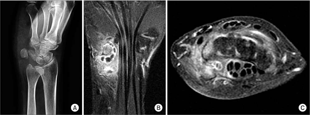
Fig. 1 (A) Preoperative oblique view showing soft tissue swelling around the pisotriquetral joint and widening of the pisotriquetral joint. (B) T2-weighted coronal magnetic resonance imaging (MRI) of the pisotriquetral joint. (C) Juxta-articular axial MRI of the pisiform demonstrating an intraarticular lobulated homogeneous material with surrounding high signal soft tissue.
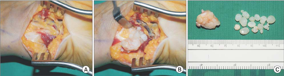
Fig. 2 (A) In Guyon's canal, the ulnar nerve is not compressed by the mass of synovial chondromatosis. (B) Intraoperative findings showing the pisiform and multiple sized cartilaginous materials of the pisotriquetral joint. A few cartilaginous bodies are attached to the synovial membrane. (C) Gross specimen after surgical excision. Small bone on the left side is the pisiform removed for the treatment of flexor carpi ulnaris tendinitis, and materials on the right side are many cartilaginous loose bodies and a few osteocartilage bodies in the pisotriquetral joint.
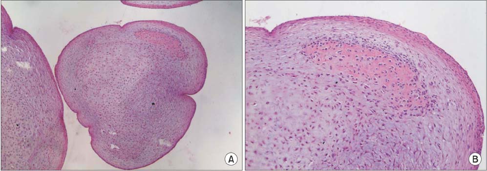
Fig. 3 Low magnification (H&E, ×10) (A) and higher magnification (H&E, ×40) (B) of the representative histology of the tumor, indicating both mature and immature cartilage formation surrounded by fibrous tissue. The beginning of mild woven bone formation is seen, but there is no evidence of malignancy.
Reference
-
1. Slesarenko YA, Hurst LC, Dagum AB. Synovial chondromatosis of the distal radioulnar joint. Hand Surg. 2004; 9(2):241–243.
Article2. Roulot E, Le Viet D. Primary synovial osteochondromatosis of the hand and wrist: report of a series of 21 cases and literature review. Rev Rhum Engl Ed. 1999; 66(5):256–266.3. Maurice H, Crone M, Watt I. Synovial chondromatosis. J Bone Joint Surg Br. 1988; 70(5):807–811.
Article4. Trias A, Quintana O. Synovial chondrometaplasia: review of world literature and a study of 18 Canadian cases. Can J Surg. 1976; 19(2):151–158.5. Villacin AB, Brigham LN, Bullough PG. Primary and secondary synovial chondrometaplasia: histopathologic and clinicoradiologic differences. Hum Pathol. 1979; 10(4):439–451.6. Norman A, Steiner GC. Bone erosion in synovial chondromatosis. Radiology. 1986; 161(3):749–752.
Article7. Kramer J, Recht M, Deely DM, et al. MR appearance of idiopathic synovial osteochondromatosis. J Comput Assist Tomogr. 1993; 17(5):772–776.
Article8. Ginaldi S. Computed tomography feature of synovial osteochondromatosis. Skeletal Radiol. 1980; 5(4):219–222.
Article9. Milgram JW. Synovial osteochondromatosis: a histopathological study of thirty cases. J Bone Joint Surg Am. 1977; 59(6):792–801.10. Kenan S, Abdelwahab IF, Klein MJ, Lewis MM. Case report 817: synovial chondrosarcoma secondary to synovial chondromatosis. Skeletal Radiol. 1993; 22(8):623–626.
