Mucosa-associated Lymphoid Tissue Lymphoma Presenting with Bowel Obstruction of the Duodenum and Small Bowels: A Case Report
- Affiliations
-
- 1Department of Radiology, Soonchunhyang University Hospital, Korea. hongses@hosp.sch.ac.kr
- KMID: 2208995
- DOI: http://doi.org/10.3348/jksr.2010.62.1.57
Abstract
- The occurrence of primary duodenal mucosa associated lymphoid tissue (MALT) lymphoma is extremely rare, and more so is the obstruction of the duodenum for the MALT lymphoma. We describe the small bowel follow through and CT findings in an uncommon case of MALT lymphoma presenting with bowel obstruction of the 2nd portion of the duodenum and small bowels.
MeSH Terms
Figure
-
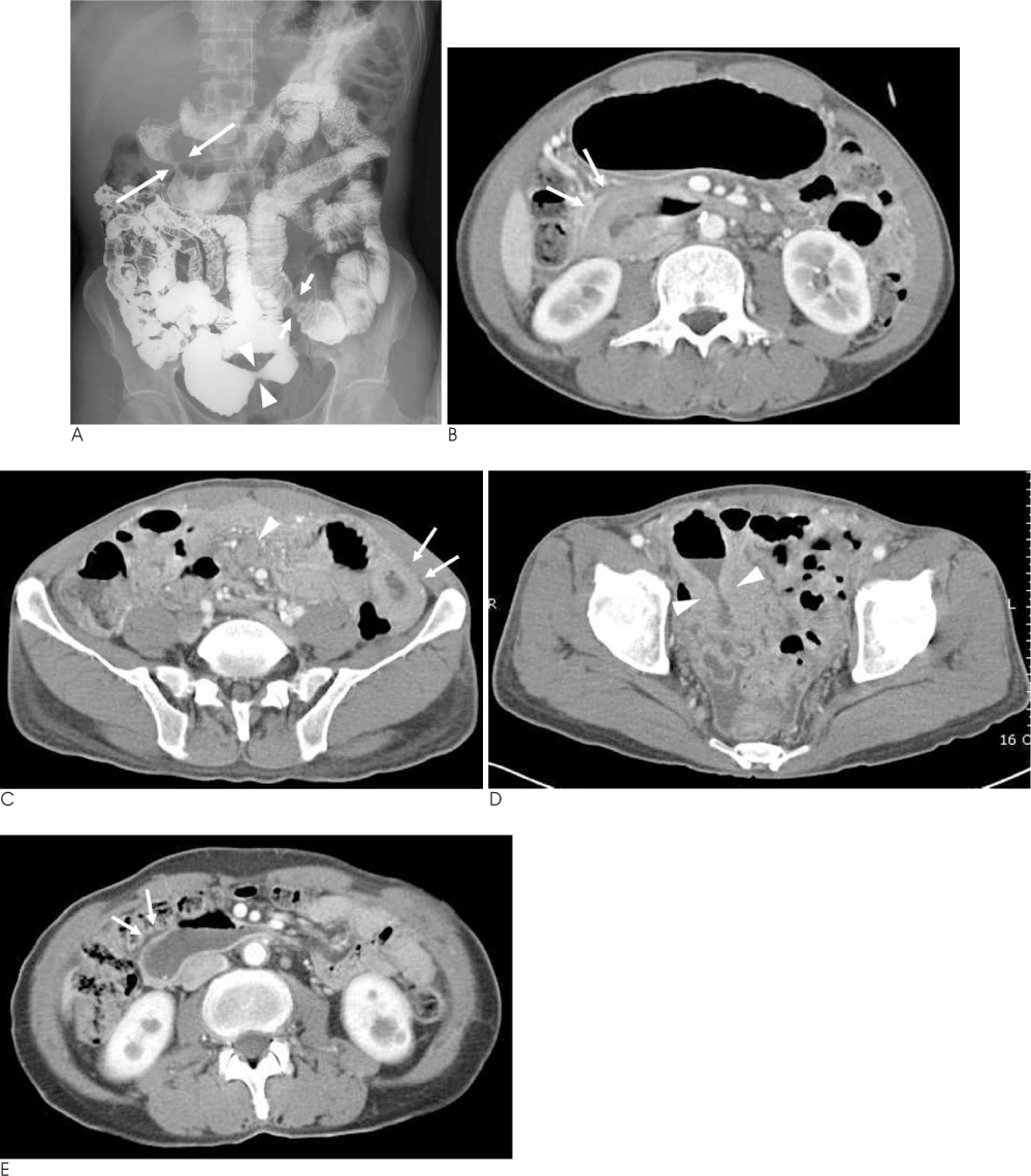
Fig. 1 56-year-old male with MALT lymphoma presenting with bowel obstruction. A. Small bowel follow through shows focal luminal narrowing (long white arrows) of the 2nd portion of the duodenum with small ulceration. And, also noted smooth luminal narrowing with fold thickening at the distal jeunal loop (short white arrows) and the pelvic ileal loop (white arrowheads). B. Contrast enhanced CT at the level of the upper abdomen shows peripheral enhancing mass-like wall thickening (white arrows) of the 2nd portion of the duodenum with proximal luminal dilatation. C. Contrast enhanced CT at the level of middle abdomen shows enhancing wall thickening (white arrows) of the proximal jejunum with multiple enlarged mesenteric lymph nodes (white arrowhead). D. Contrast enhanced CT scan at the level of lower abdomen shows dumbell-shape luminal narrowing (white arrowheads) of the pelvic ileal loop. E. Follow-up CT after 10 months of chemotherapy shows improvement of wall thickening (white arrows) of the duodenum and small bowel loops. Multiple mesenteric lymph nodes are also improved (not shown).
Reference
-
1. Isaacson PG, Wright DH. Malignant lymphoma of mucosa-associated lymphoid tissue. A distinctive type of B-cell lymphoma. Cancer. 1983; 52:1410–1416.2. Tadmor T, Rainis T, Bejar J, Attias D, Lavy A. Primary duodenal mucosa-associated lymphoid tissue (MALT) lymphoma a rare presentation of gastric outlet obstruction. Can J Gastroenterol. 2007; 21:393–395.3. Patel VG, Eltayeb OM, Henderson VJ, Lyons R, Martin D, Hamami A, et al. Primary duodenal low-grade mucosa-associated lymphoid tissue lymphoma presenting with outlet obstruction. Am Surg. 2004; 70:613–616.4. Parsonnet J, Hansen S, Rodriguez L, Gelb AB, Warnke RA, Jellum E, et al. Helicobacter pylori infection and gastric lymphoma. N Engl J Med. 1994; 330:1267–1271.5. Yoo CC, Levine MS, Furth EE, Salhany KE, Rubesin SE, Laufer I, et al. Gastric mucosa-associated lymphoid tissue lymphoma: radiographic findings in six patients. Radiology. 1998; 208:239–243.6. Kim YH, Lim HK, Han JK, Choi BI, Kim YI, Lee WJ, et al. Lowgrade gastric mucosa-associated lymphoid tissue lymphoma: correlation of radiographic and pathologic findings. Radiology. 1999; 212:241–248.7. Fischbach W, Dragosics B, Kolve-Goebeler ME, Ohmann C, Greiner A, Yang Q, et al. Primary gastric B-cell lymphoma: results of a prospective multicenter study. The German-Austrian gastrointestinal lymphoma study group. Gastroenterology. 2000; 119:1191–1202.8. Nahm DI, Baek IH, Lee MS. Primary duodenal MALT lymphoma. Korean J Gastroenterol. 2007; 49:343–345.9. Du MQ, Xu CF, Diss TC, Peng HZ, Wotherspoon AC, Isaacson PG, et al. Intestinal dissemination of gastric mucosa-associated lymphoid tissue lymphoma. Blood. 1996; 88:4445–4451.10. Nebiki H, Harihara S, Tsukuda H, Inoue T, Arakawa T. Regression of gastric MALT lymphoma after unsuccessful anti-H. pylori therapy. Am J Gastroenterol. 2000; 95:3684–3686.
- Full Text Links
-
- Actions
-
Cited
- CITED
-
- Close
- Share
-
- Similar articles
-
- A Case of Gastroduodenal Mucosa-associated Lymphoid Tissue Lymphoma Regression after Eradication of Helicobacter pylori
- A Case of Simultaneous Primary Gastric and Duodenal Mucosa-Associated Lymphoid Tissue Lymphoma after Therapeutic Endoscopy
- A Case of Gastroduodenal Mucosa Associated Lymphoid Tissue Lymphoma Regressed After Helicobacter pylori Eradication
- Duodenal Mucosa-associated Lymphoid Tissue Lymphoma Treated with Chemotherapy
- A Case of Ileal Mucosa-associated Lymphoid Tissue Lymphoma Accompanied by Luminal Stricture and Arterial Spurting
