J Korean Soc Radiol.
2010 Mar;62(3):207-210. 10.3348/jksr.2010.62.3.207.
Circumaortic Left Brachiocephalic Vein: CT Findings
- Affiliations
-
- 1Department of Radiology, Sanggye Paik Hospital, Inje University College of Medicine, Korea. S2621@paik.ac.kr
- KMID: 2208914
- DOI: http://doi.org/10.3348/jksr.2010.62.3.207
Abstract
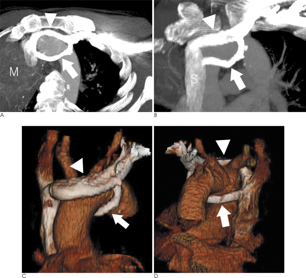
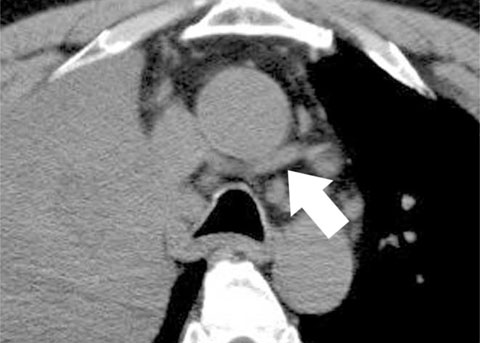
- The left brachiocephalic vein usually passes superior and anterior to the aortic arch. In rare cases, this vein follows an anomalous course; for example, a circumaortic left brachiocephalic vein. We present the case of a circumaortic left brachiocephalic vein in a 53-year-old male with lung cancer. A multidetector CT showed that the two branches of the left brachiocephalic vein circumscribed the aortic arch. The anterior branch was above and the posterior branch was below the aortic arch. Both branches drained into the superior vena cava.
MeSH Terms
Figure
Reference
-
1. Standring S. Gray's anatomy. 39th. Edinburgh: Elsevier Churchil Livingstone;2005. p. 1027.2. Kerschner L. Zur. Morphologie der Vena cava inferior. Anat Anz. 1888; 3:808–823.3. Takada Y, Narimatsu A, Kohno A, Kawai C, Hara H, Harasawa A, et al. Anomalous left brachiocephalic vein: CT findings. J Comput Assist Tomogr. 1992; 16:893–896.4. Gerlis LM, Ho SY. Anomalous subaortic position of the brachiocephalic (innominate) vein: a review of published reports and report of three new cases. Br Heart J. 1989; 61:540–545.5. Arey LB. The vascular system. In : Arey LB, editor. Developmental anatomy. 7th. Philadelphia: Saunders;1966. p. 342–374.6. Maxwell DS. Embryology of the vascular system. In : Moore WS, editor. Vascular surgery: a comprehensive reviews. Philadelphia: Saunders;1991. p. 20–36.7. Kellman GM, Alpern MB, Sandler MA, Craig BM. Computed tomography of vena caval anomalies with embryologic correlation. Radiographics. 1988; 8:533–556.8. Adachi B. Das venensystem der Japaner. Anatomie der Japaner II. Tokyo: Kenkyusha;1933. p. 83–87.9. Yigit AE, Haliloglu M, Karcaaltincaba M, Ariyurek MO. Retrotracheal aberrant left brachiocephalic vein: CT findings. Pediatr Radiol. 2008; 38:322–324.10. Choi JY, Jung MJ, Kim YH, Noh CI, Yun YS. Anomalous subaortic position of the brachiocephalic vein (innominate vein): an echocardiographic study. Br Heart J. 1990; 64:385–387.
- Full Text Links
- Actions
-
Cited
- CITED
-
- Close
- Share
- Similar articles
-
- Double Left Brachiocephalic Veins with Persistent Left Superior Vena Cava: A Case Report
- MDCT Findings of Right Circumaortic Renal Vein with Ectopic Kidney
- Subaortic Left Brachiocephalic Vein
- The Anomalous Left Brachiocephalic Vein in Adults
- An Unusual Cause of Left Brachiocephalic Vein Occlusion: Extrinsic Compression by the Aortic Arch in a Hemodialysis Patient


