Double Left Brachiocephalic Veins with Persistent Left Superior Vena Cava: A Case Report
- Affiliations
-
- 1Department of Radiology, Yeungnam University College of Medicine, Daegu, Korea. stallin64@naver.com
- KMID: 1819779
- DOI: http://doi.org/10.3348/jksr.2014.71.2.55
Abstract
- Anomalous left brachiocephalic vein (ALBCV) is a rare condition of the major thoracic veins. It is usually associated with a congenital cardiac anomaly. Most reports on ALBCV are on aberrant left BCV, and there are few reports on double left BCV. Persistent left superior vena cava (PLSVC) is also a rare vascular anomaly that is caused by the failure of the left anterior cardinal vein to regress. To our knowledge, double left BCV with PLSVC has not been reported. Here, we report a case of double left BCV with PLSVC in a 72-year-old male patient with no previous cardiac abnormality.
Figure
-
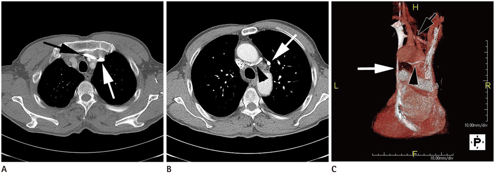
Fig. 1 Double left brachiocephalic veins (BCV) with persistent left superior vena cava (PLSVC) in a 72-year-old male patient. A. Axial image of the contrast-enhanced CT scan at the level above the aortic arch, showing that the normally placed branch of the left BCV (black arrow) courses obliquely anterior to the ascending aorta. PLSVC is also noted (white arrow). B. At the level of the ascending aorta, subaortic branch of the left BCV (arrowhead) showing the obliquely downward course behind the ascending aorta and drainage into the right SVC. PLSVC is also noted (white arrow). C. Three-dimensional reconstruction images of the PA view revealing two connections between the two SVCs through the double left BCV. Black arrow = normally placed branch of the left BCV, white arrow = PLSVC, arrowhead = subaortic branch of the left BCV.
Reference
-
1. Chern MS, Ko JS, Tsai A, Wu MH, Teng MM, Chang CY. Aberrant left brachiocephalic vein: CT imaging findings and embryologic correlation. Eur Radiol. 1999; 9:1835–1839.2. Chen SJ, Liu KL, Chen HY, Chiu IS, Lee WJ, Wu MH, et al. Anomalous brachiocephalic vein: CT, embryology, and clinical implications. AJR Am J Roentgenol. 2005; 184:1235–1240.3. Takada Y, Narimatsu A, Kohno A, Kawai C, Hara H, Harasawa A, et al. Anomalous left brachiocephalic vein: CT findings. J Comput Assist Tomogr. 1992; 16:893–896.4. Steinberg I, Dubilier W Jr, Lukas DS. Persistence of left superior vena cava. Dis Chest. 1953; 24:479–488.5. Subirana MT, de Leval M, Somerville J. Double left innominate vein: an unusual cross-sectional echocardiographic appearance. Int J Cardiol. 1986; 12:263–265.6. Nandy K, Blair CB Jr. Double superior venae cabae with completely paired azygos veins. Anat Rec. 1965; 151:1–9.7. Starck D. Embryologie. Ein Lehrbuch auf allgemein biologischer Grundlage. 3rd ed. Stuttgart: Geor Thieme;1975.8. Webb WR, Gamsu G, Speckman JM, Kaiser JA, Federle MP, Lipton MJ. Computed tomographic demonstration of mediastinal venous anomalies. AJR Am J Roentgenol. 1982; 139:157–161.
- Full Text Links
-
- Actions
-
Cited
- CITED
-
- Close
- Share
-
- Similar articles
-
- A Case of Persistent Left Superior Vena Cava with Interruption of Inferior Vena Cava
- Persistent Left Superior Vena Cava with Absent Right Superior Vena Cava and Large Atrial Septal Defect in Visceroatrial Situs solitus
- A Case of Persistent Left Superior Vena Cava Detected on Fetal Echocardiography
- A Case of Behcet's Disease with Superior Vena Cava Syndrome
- Congenital Absence of the Azygos Vein with Persistent Left Superior Vena Cava: A Case Report
