J Korean Soc Radiol.
2013 Aug;69(2):89-92. 10.3348/jksr.2013.69.2.89.
Regional Cortical Hyperperfusion on Perfusion CT during Postictal Motor Deficit: A Case Report
- Affiliations
-
- 1Department of Radiology, Haeundae Paik Hospital, Inje University College of Medicine, Busan, Korea. sartre81@gmail.com
- KMID: 2208810
- DOI: http://doi.org/10.3348/jksr.2013.69.2.89
Abstract
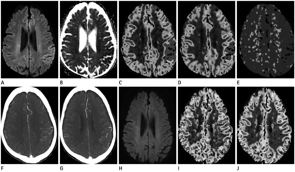
- Postictal neurologic deficit is a well-known complication mimicking the manifestation of a stroke. We present a case of a patient with clinical evidence of Todd's paralysis correlating with reversible postictal parenchymal changes on perfusion CT and magnetic resonance (MR) imaging. In this case, perfusion CT and MR imaging were helpful in the differential diagnosis of stroke-mimicking conditions.
MeSH Terms
Figure
Reference
-
1. Davis DP, Robertson T, Imbesi SG. Diffusion-weighted magnetic resonance imaging versus computed tomography in the diagnosis of acute ischemic stroke. J Emerg Med. 2006; 31:269–277.2. Mathews MS, Smith WS, Wintermark M, Dillon WP, Binder DK. Local cortical hypoperfusion imaged with CT perfusion during postictal Todd’s paresis. Neuroradiology. 2008; 50:397–401.3. Masterson K, Vargas MI, Delavelle J. Postictal deficit mimicking stroke: role of perfusion CT. J Neuroradiol. 2009; 36:48–51.4. Hemmen TM, Meyer BC, McClean TL, Lyden PD. Identification of nonischemic stroke mimics among 411 code strokes at the University of California, San Diego, Stroke Center. J Stroke Cerebrovasc Dis. 2008; 17:23–25.5. Weinand ME, Carter LP, el-Saadany WF, Sioutos PJ, Labiner DM, Oommen KJ. Cerebral blood flow and temporal lobe epileptogenicity. J Neurosurg. 1997; 86:226–232.6. Moritani T, Smoker WR, Sato Y, Numaguchi Y, Westesson PL. Diffusion-weighted imaging of acute excitotoxic brain injury. AJNR Am J Neuroradiol. 2005; 26:216–228.7. Warach S, Levin JM, Schomer DL, Holman BL, Edelman RR. Hyperperfusion of ictal seizure focus demonstrated by MR perfusion imaging. AJNR Am J Neuroradiol. 1994; 15:965–968.8. Chan S, Chin SS, Kartha K, Nordli DR, Goodman RR, Pedley TA, et al. Reversible signal abnormalities in the hippocampus and neocortex after prolonged seizures. AJNR Am J Neuroradiol. 1996; 17:1725–1731.9. Wiest R, von Bredow F, Schindler K, Schauble B, Slotboom J, Brekenfeld C, et al. Detection of regional blood perfusion changes in epileptic seizures with dynamic brain perfusion CT--a pilot study. Epilepsy Res. 2006; 72:102–110.10. Szabo K, Poepel A, Pohlmann-Eden B, Hirsch J, Back T, Sedlaczek O, et al. Diffusion-weighted and perfusion MRI demonstrates parenchymal changes in complex partial status epilepticus. Brain. 2005; 128(Pt 6):1369–1376.
- Full Text Links
- Actions
-
Cited
- CITED
-
- Close
- Share
- Similar articles
-
- Acetazolamide-Challenged Brain CT Perfusion before and after Carotid Stenting
- Cerebral hyperperfusion syndrome after endovascular stent graft reconstruction for postirradiated carotid blowout syndrome: a case report
- Segmental abnormal perfusion in the liver: Relation between hepatic arterial and portal vein blood flow inn the fast contrast CT
- MR Findings of Seizure-Related Cerebral Cortical Lesions during Periictal Period
- Hyperperfusion in DWI Abnormality in a Patient with Acute Symptomatic Hypoglycemic Encephalopathy


