J Korean Soc Radiol.
2013 Sep;69(3):177-179. 10.3348/jksr.2013.69.3.177.
Disorganized Foliation of Unilateral Cerebellar Hemisphere as Cerebellar Cortical Dysplasia in Patients with Recurrent Seizures: A Case Report
- Affiliations
-
- 1Department of Radiology, Haeundae Paik Hospital, Inje University College of Medicine, Busan, Korea. sartre81@gmail.com
- KMID: 2208797
- DOI: http://doi.org/10.3348/jksr.2013.69.3.177
Abstract
- We present a rare case of abnormal foliation for one cerebellar hemisphere on MR imaging, showing vertically-oriented folia. Foliation of contralateral cerebellar hemisphere and other structures in the posterior fossa were normal, and the patient has no neurologic deficits. This rare and unique abnormality is considered a kind of developmental error of the cerebellum.
MeSH Terms
Figure
-
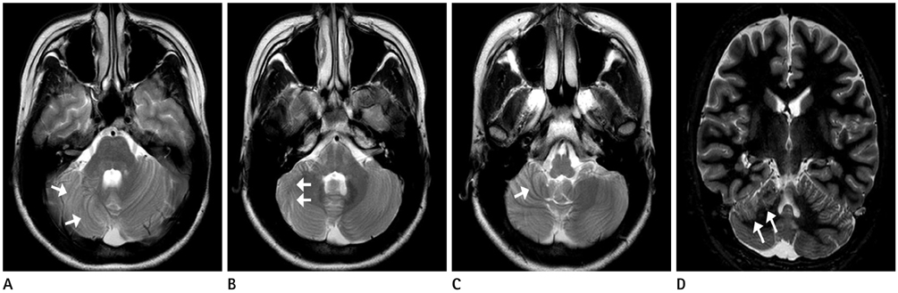
Fig. 1 A 26-year-old woman with recurrent generalized tonic-clonic seizures. A-C. Axial T2-weighted images through the lower midbrain to the medulla oblongata. Folia and fissures of the right cerebellar hemisphere show vertically oriented (arrows), and it is most prominent in the inferior aspect. The folia of the upper aspect of the ipsilateral hemisphere run almost horizontally. The right cerebellar hemisphere is smaller than left one. Foliation of the left cerebral hemisphere and configuration of the vermis and fourth ventricle are normal. D. On coronal T2-weighted images, the corticomedullary junction of the affected cerebellar hemisphere is irregular and nodular configurations with indistinct margin (arrows).
Reference
-
1. Aida N, Tamagawa K, Takada K, Yagishita A, Kobayashi N, Chikumaru K, et al. Brain MR in Fukuyama congenital muscular dystrophy. AJNR Am J Neuroradiol. 1996; 17:605–613.2. Barkovich AJ. Neuroimaging manifestations and classification of congenital muscular dystrophies. AJNR Am J Neuroradiol. 1998; 19:1389–1396.3. Demaerel P, Lagae L, Casaer P, Baert AL. MR of cerebellar cortical dysplasia. AJNR Am J Neuroradiol. 1998; 19:984–986.4. Sasaki M, Oikawa H, Ehara S, Tamakawa Y, Takahashi S, Tohgi H. Disorganised unilateral cerebellar folia: a mild form of cerebellar cortical dysplasia? Neuroradiology. 2001; 43:151–155.5. Soto-Ares G, Delmaire C, Deries B, Vallee L, Pruvo JP. Cerebellar cortical dysplasia: MR findings in a complex entity. AJNR Am J Neuroradiol. 2000; 21:1511–1519.6. Yachnis AT, Rorke LB, Trojanowski JQ. Cerebellar dysplasias in humans: development and possible relationship to glial 179and primitive neuroectodermal tumors of the cerebellar vermis. J Neuropathol Exp Neurol. 1994; 53:61–71.7. Aida N, Yagishita A, Takada K, Katsumata Y. Cerebellar MR in Fukuyama congenital muscular dystrophy: polymicrogyria with cystic lesions. AJNR Am J Neuroradiol. 1994; 15:1755–1759.8. Barth PG. Pontocerebellar hypoplasias. An overview of a group of inherited neurodegenerative disorders with fetal onset. Brain Dev. 1993; 15:411–442.9. Larroche JC. Morphological criteria of central nervous system development in the human foetus. J Neuroradiol. 1981; 8:93–108.10. Takanashi J, Sugita K, Barkovich AJ, Takano H, Kohno Y. Partial midline fusion of the cerebellar hemispheres with vertical folia: a new cerebellar malformation? AJNR Am J Neuroradiol. 1999; 20:1151–1153.
- Full Text Links
-
- Actions
-
Cited
- CITED
-
- Close
- Share
-
- Similar articles
-
- Bilateral Cerebellar Cortical Dysplasia without Other Malformations: A Case Report
- A Cerebellar Infarction Presented with a Clinical Seizure
- Cerebellar Cortical Artery Dissection Technique for the Preservation of Operative Fields during Microvascular Decompression for Hemifacial Spasm: Technical Note
- Transient Cerebellar Mutism after Total Removal of Medulloblastoma in a Child: Case Report
- Gliomatosis Cerebri in the Brain Stem and Unilateral Cerebellar Hemisphere: Case Report
