Clinical Experience of 3T Breast MRI in Detecting the Additional Lesions in Breast Cancer Patients
- Affiliations
-
- 1Department of Radiology, College of Medicine, The Catholic University of Korea, Korea. rad-ksh@catholic.ac.kr
- 2Department of Hospital Pathology, College of Medicine, The Catholic University of Korea, Korea.
- KMID: 2206909
- DOI: http://doi.org/10.13104/jksmrm.2010.14.2.121
Abstract
- PURPOSE
The purpose of this study was to evaluate the diagnostic accuracy of 3.0-T breast MRI for detecting additional breast cancer soon after the initial diagnosis of breast cancer.
MATERIALS AND METHODS
From March to June 2009, 101 patients recently diagnosed breast cancer underwent breast MRI and surgery. Parameters analyzed on MRI were total extent of tumor, suspicious findings of multifocal, multicentric, or contralateral cancer. The diagnosis of MRI-detected cancer was confirmed by means of biopsy or surgical specimen evaluation after the localization.
RESULTS
MRI showed 37 additional suspicious findings in 34 patients. Twenty nine findings were true-positive (29/37, 78.4%), including 16 cases of multifocality, 11 cases of multicentricity and 2 cases of contralateral cancer. Among these cancers, 13 (44.8%) were ductal carcinoma in situ (DCIS) and 16 (55.1%) were infiltrating cancer. Eight findings were false-positive (8/37, 21.6%) including 6 cases of benign disease and 2 cases of high-risk lesions.
CONCLUSION
In women with recently diagnosed breast cancer, 3.0-T MR imaging showed additional suspicious findings in 33.7%. The sensitivity and specificity for detecting additional breast cancer was 100% and 89.3%, respectively.
Keyword
MeSH Terms
Figure
-
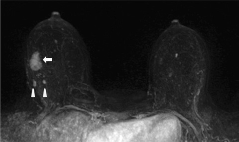
Fig. 1 35-year-old woman with invasive ductal cancer and true positive multifocal malignancies. 3D MIP image shows an index mass (arrow) and two multifocal daughter nodules (arrowheads) at upper outer quadrant of right breast.
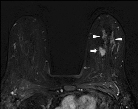
Fig. 2 53-year-old woman with invasive ductal cancer and true positive multicentric malignancies. Axial subtraction image shows an index mass (arrow) at upper inner quadrant of left breast. Also detected multifocal and multicentric malignancies (arrowheads) are noted at left upper breast.

Fig. 3 63-year-old woman with invasive ductal cancer and true positive contralateral malignancy. Axial subtraction image shows an index mass at right breast and an irregular spiculated homogeneously enhancing mass at left breast.
Reference
-
1. Elsamaloty H, Elzawawi MS, Mohammad S, Herial N. Increasing accuracy of detection of breast cancer with 3-T MRI. AJR Am J Roentgenol. 2009. 192:1142–1148.2. Orel SG, Schnall MD. MR imaging of the breast for the detection, diagnosis, and staging of breast cancer. Radiology. 2001. 220:13–30.3. Riedl CC, Ponhold L, Flory D, et al. Magnetic resonance imaging of the breast improves detection of invasive cancer, preinvasive cancer, and premalignant lesions during surveillance of women at high risk for breast cancer. Clin Cancer Res. 2007. 13:6144–6152.4. Kuhl CK. Breast MR imaging at 3T. Magn Reson Imaging Clin N Am. 2007. 15:315–320.5. Sasaki M, Shibata E, Kanbara Y, Ehara S. Enhancement effects and relaxivities of gadolinium-DTPA at 1.5 versus 3 Tesla: a phantom study. Magn Reson Med Sci. 2005. 4:145–149.6. Liberman L, Morris EA, Dershaw DD, Abramson AF, Tan LK. MR imaging of the ipsilateral breast in women with percutaneously proven breast cancer. AJR Am J Roentgenol. 2003. 180:901–910.7. Mameri CS, Kemp C, Goldman SM, Sobral LA, Ajzen S. Impact of breast MRI on surgical treatment, axillary approach, and systemic therapy for breast cancer. Breast J. 2008. 14:236–244.8. Schmitz AC, Peters NH, Veldhuis WB, et al. Contrast-enhanced 3.0-T breast MRI for characterization of breast lesions: increased specificity by using vascular maps. Eur Radiol. 2008. 18:355–364.9. Kuhl CK, Jost P, Morakkabati N, Zivanovic O, Schild HH, Gieseke J. Contrast-enhanced MR imaging of the breast at 3.0 and 1.5 T in the same patients: initial experience. Radiology. 2006. 239:666–676.10. Wiener JI, Schilling KJ, Adami C, Obuchowski NA. Assessment of suspected breast cancer by MRI: a prospective clinical trial using a combined kinetic and morphologic analysis. AJR Am J Roentgenol. 2005. 184:878–886.11. Kriege M, Brekelmans CT, Boetes C, et al. Efficacy of MRI and mammography for breast-cancer screening in women with a familial or genetic predisposition. N Engl J Med. 2004. 351:427–437.12. Leach MO, Boggis CR, Dixon AK, et al. Screening with magnetic resonance imaging and mammography of a UK population at high familial risk of breast cancer: a prospective multicentre cohort study (MARIBS). Lancet. 2005. 365:1769–1778.
- Full Text Links
-
- Actions
-
Cited
- CITED
-
- Close
- Share
-
- Similar articles
-
- Clinical Outcome of Magnetic Resonance Imaging-Detected Additional Lesions in Breast Cancer Patients
- Low Rates of Additional Cancer Detection by Magnetic Resonance Imaging in Newly Diagnosed Breast Cancer Patients Who Undergo Preoperative Mammography and Ultrasonography
- Magnetic Resonance Findings of Breast Diseases
- The Clinical Significance of Preoperative MRI for Determination of Surgery in Breast Cancer
- Usefulness of Preoperative Breast MRI in Breast Cancer Patients Diagnosed with Tumor Removal Using a US-guided Mammotome
