J Korean Rheum Assoc.
2009 Jun;16(2):167-169. 10.4078/jkra.2009.16.2.167.
Localized Cutaneous Sclerosis Presenting as Whitish Guttate Spots
- Affiliations
-
- 1Department of Dermatology, Hanyang University College of Medicine, Seoul, Korea. cwlee@hanyang.ac.kr
- KMID: 2202134
- DOI: http://doi.org/10.4078/jkra.2009.16.2.167
Abstract
- No abstract available.
MeSH Terms
Figure
-
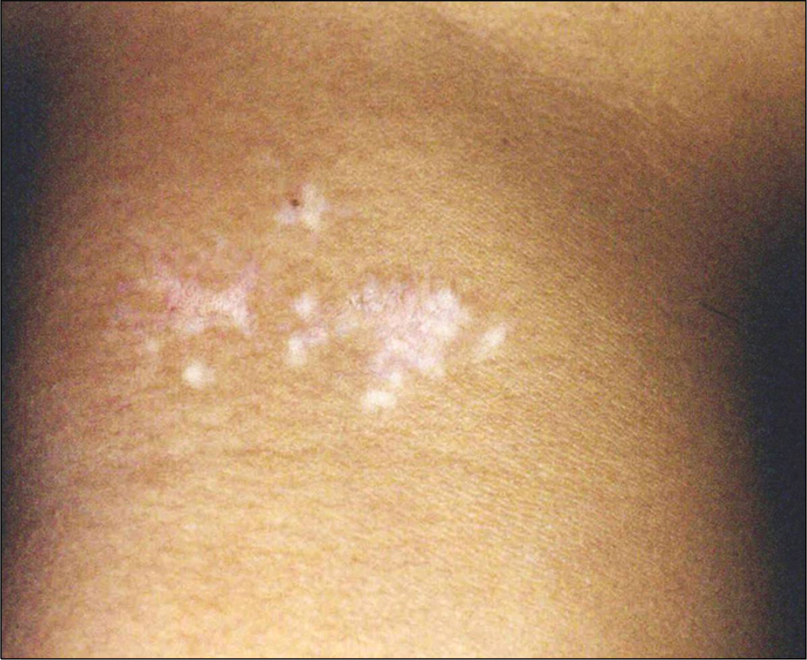
Fig. 1. Localized ill-defined whitish-brown sclerotic patches on the right neck.
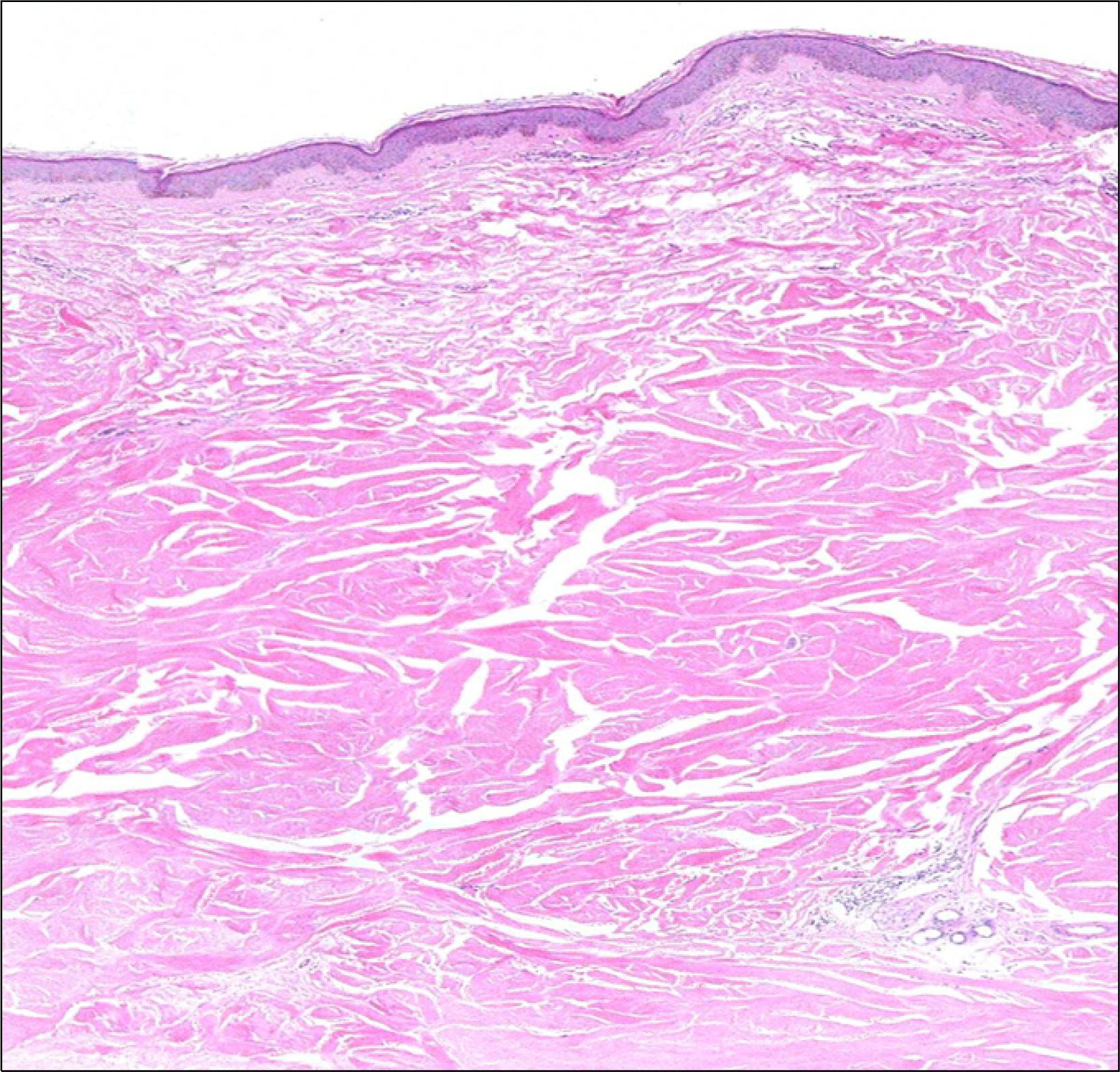
Fig 2. Biopsy specimen showing pigment incontinence and sclerotic collagen bundles with atrophy of the eccrine glands (H&E, ×40).
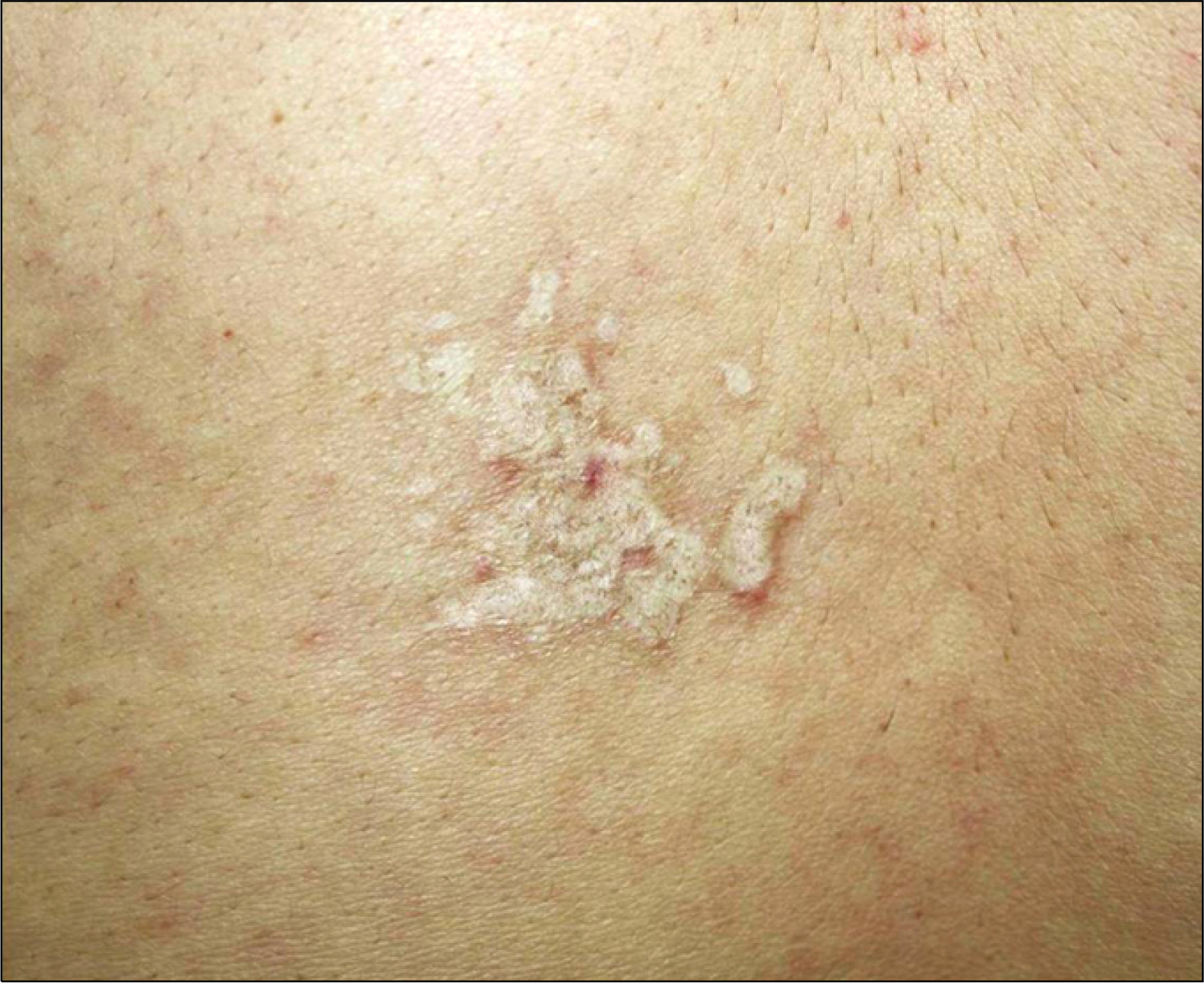
Fig. 3. Localized whitish-yellow confluent and guttated sclerotic plaque and patches on the lower back.
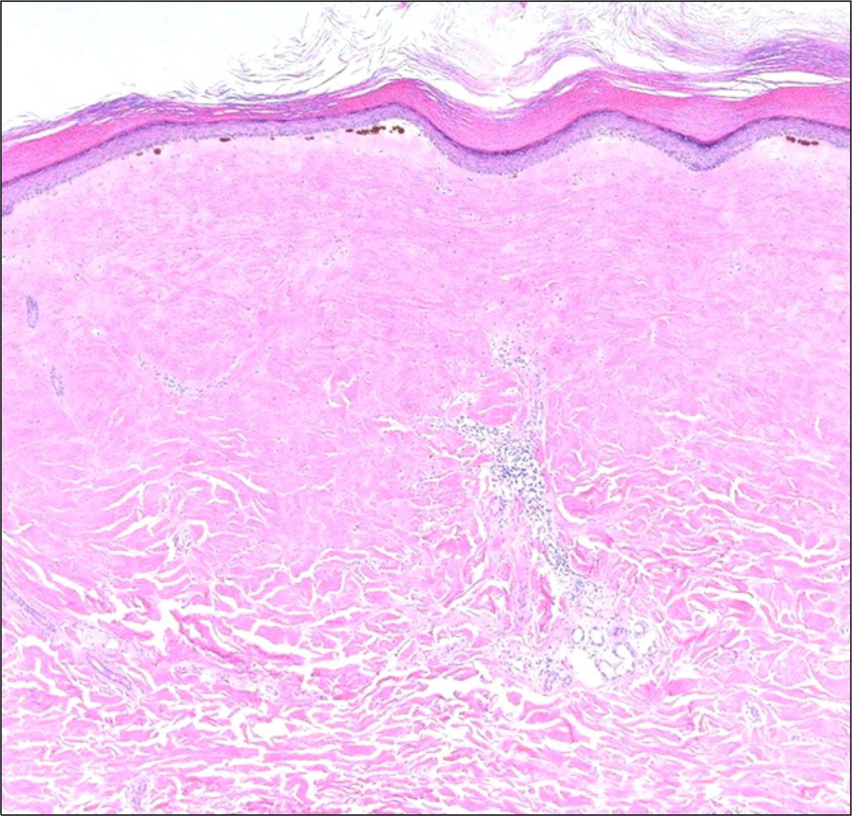
Fig. 4. Loss of epidermal melanocyte (with some melano-phages) and closely packed collagen bundles, with entrapped skin appendages are observed (H&E, ×40).
Reference
-
References
1. Laxer RM, Zulian F. Localized scleroderma. Curr Opin Rheumatol. 2006; 18:606–13.
Article2. Peterson LS, Nelson AM, Daniel WP. Classification of morphea (Localized scleroderma). Mayo Clin Proc. 1995; 70:1068–76.
Article3. Goodfield MJD, Jones SK, Veale DJ. The ‘connective tissue disease'. Tony B, Stephen B, Neil C, Christopher G, editors. Rook's textbook of dermatology. 7th ed.p. 56.70–56.80, Oxford, Blackwell publishing;2004.
