J Korean Rheum Assoc.
2010 Dec;17(4):340-347. 10.4078/jkra.2010.17.4.340.
MR Imaging of Ankylosing Spondylitis
- Affiliations
-
- 1Department of Radiology, Yeouido St. Mary's Hospital, The Catholic University of Korea, Seoul, Korea.
- 2Department of Radiology, Seoul St. Mary's Hospital, The Catholic University of Korea, Seoul, Korea. whjee@catholic.ac.kr
- KMID: 2201891
- DOI: http://doi.org/10.4078/jkra.2010.17.4.340
Abstract
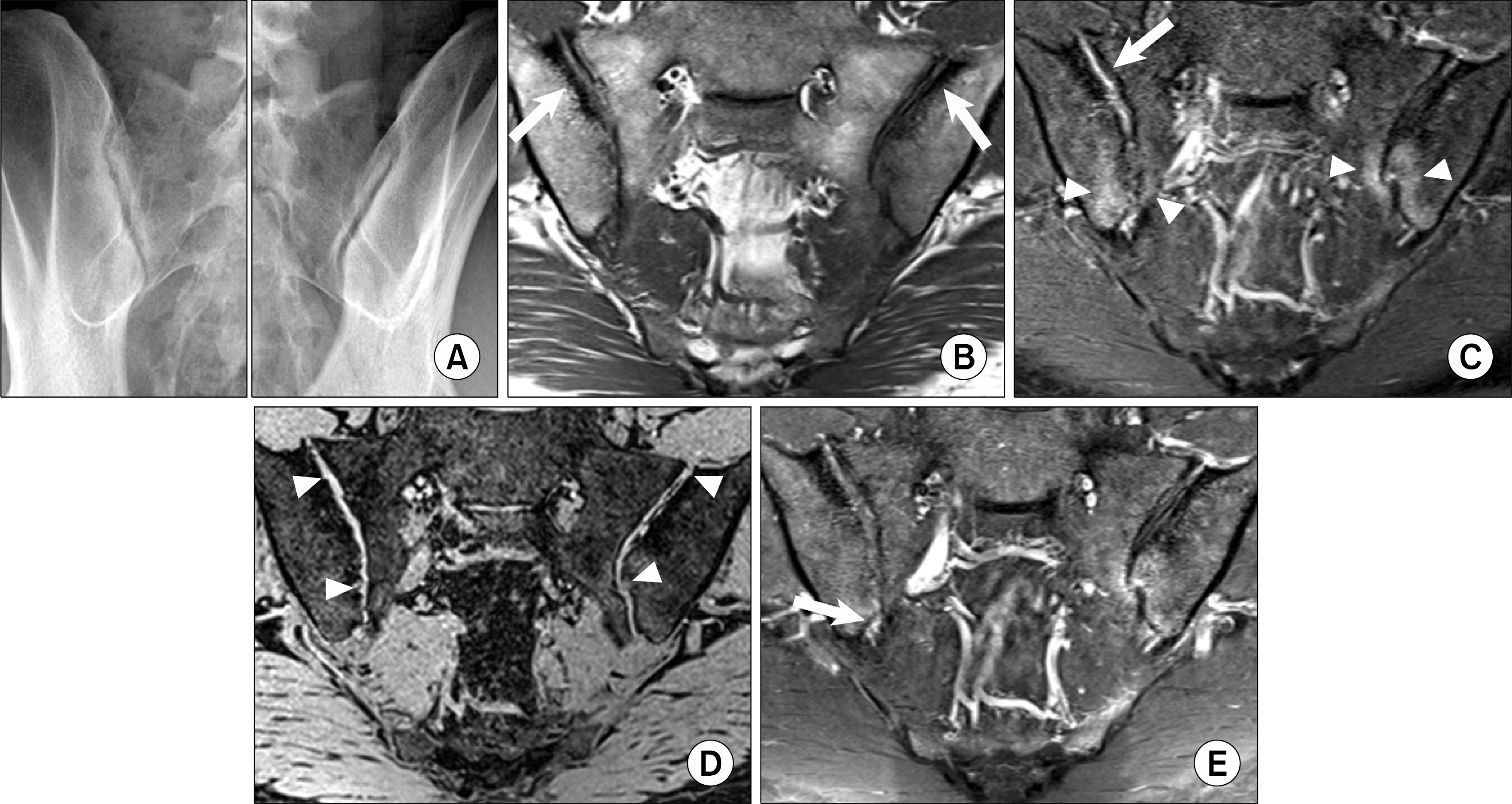
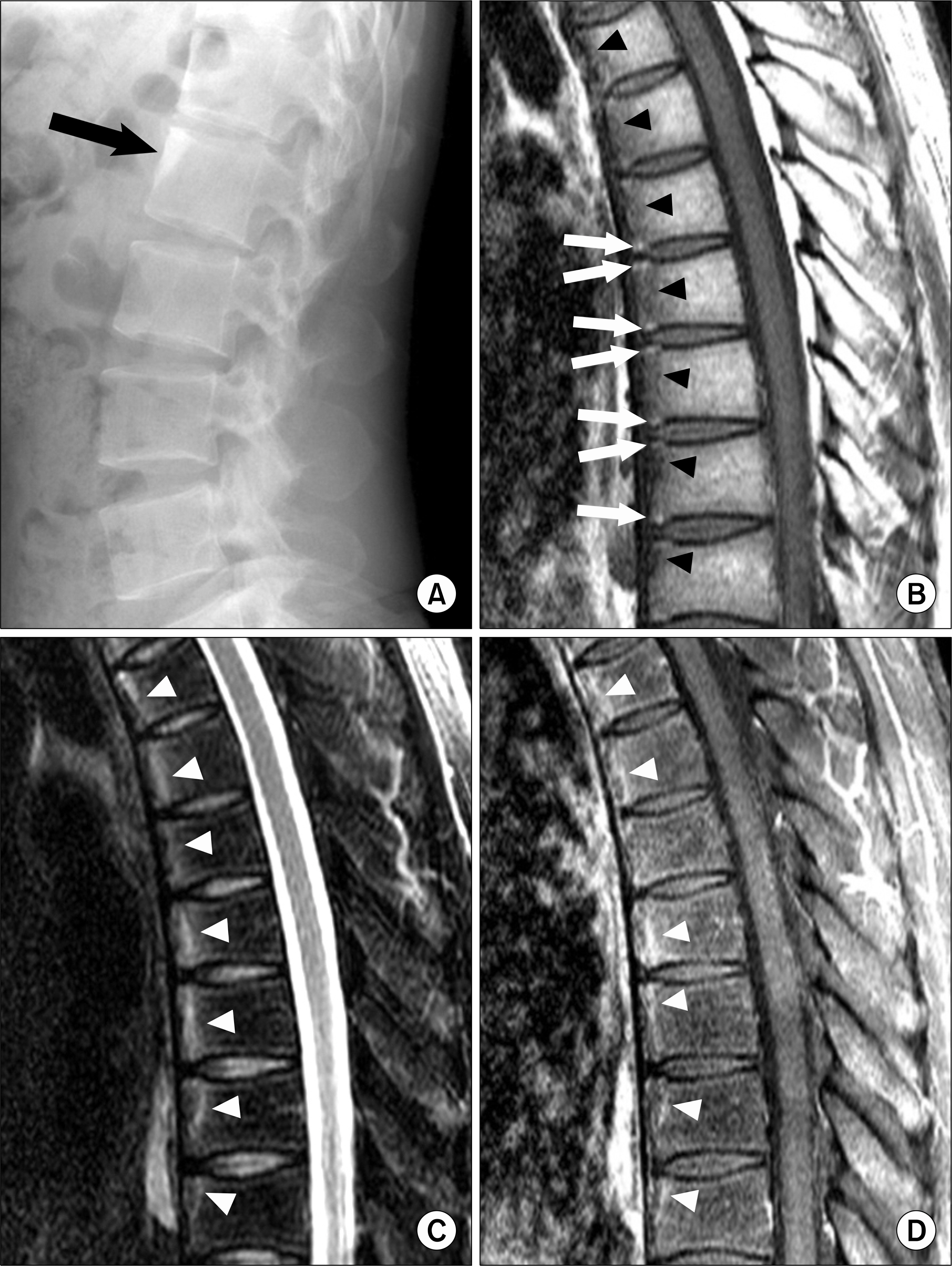
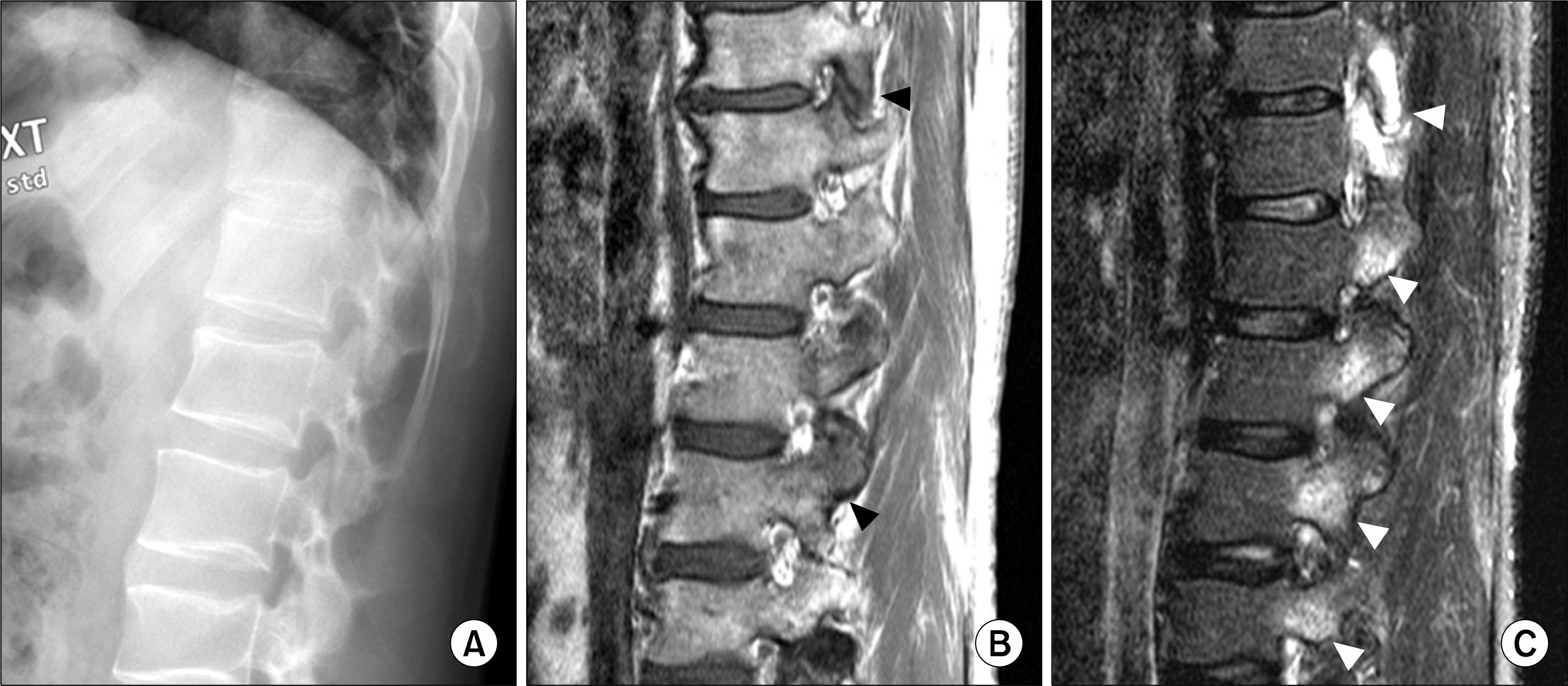
- Magnetic resonance imaging (MRI) is a highly reliable tool for diagnosing ankylosing spondylitis. MRI can identify cartilage abnormalities, subcortical erosions, bone marrow edema with inflammation, and synovial enhancement. Subchondral sclerosis and juxta-articular fat deposition are noted in the chronic stage of ankylosing spondylitis. Spinal changes associated with spondyloarthropathy are florid anterior spondylitis (or Romanus lesion), florid diskitis (Anderson lesion), ankylosis, and arthritis of the apophyseal and costovertebral joints. A MRI grading system for inflammation in sacroiliac joints and the spine could help clinicians evaluate the anti-inflammatory efficacy of therapeutics. Newer technologies based on MRI are aimed at broadening the diagnostic scope and facilitating the quantification of active inflammation but still require extensive validation.
MeSH Terms
Figure
Reference
-
1). Ryan LM., Carrera GF., Lightfoot RW Jr., Hoffman RG., Kozin F. The radiographic diagnosis of sacroiliitis: a comparison of different views with computed tomograms of the sacroiliac joints. Arthritis Rheum. 1983. 26:760–3.2). Fam AG., Rubenstein JD., Chin Sangh H., Leung FY. Computed tomography in the diagnosis of early ankylosing spondylitis. Arthritis Rheum. 1985. 28:930–7.
Article3). Dequeker J., Godderis T., Walravens M., De Roo M. Evaluation of sacroiliitis: comparison of radiological and radionuclide techniques. Radiology. 1978. 128:687–9.
Article4). Blum U., Buitrago-Tellez C., Mundinger A., Krause T., Laubenberger J., Vaith P, et al. Magnetic resonance imaging (MRI) for detection of active sacroiliitisn - a prospective study comparing conventional radiography, scintigraphy, and contrast enhanced MRI. J Rheumatol. 1996. 23:2107–15.5). Braun J., Bollow M., Eggens U., KoUnig H., Distler A., Sieper J. Use of dynamic magnetic resonance imaging with fast imaging in the detection of early and advanced sacroiliitis in spondyloarthropathy patients. Arthritis Rheum. 1994. 37:1039–45.6). Braun J., Bollow M., Sieper J. Radiologic diagnosis and pathology of the spondyloarthropathies. Rheum Dis Clin North Am. 1998. 24:697–735.
Article7). Baraliakos X., LandeweA R., Hermann KG., Listing J., Golder W., Brandt J, et al. Inflammation in ankylosing spondylitis: a systematic description of the extent and frequency of acute spinal changes using magnetic resonance imaging. Ann Rheum Dis. 2005. 64:730–4.
Article8). Hermann KG., Althoff CE., Schneider U., ZuUhlsdorf S., Lembcke A., Hamm B, et al. Spinal changes in patients with spondyloarthritis: comparison of MR imaging and radiographic appearances. Radiographics. 2005. 25:559–70.
Article9). Kim NR., Choi JY., Hong SH., Jun WS., Lee JW., Choi JA, et al. “MR corner sign”: value for predicting presence of ankylosing spondylitis. AJR Am J Roentgenol. 2008. 191:124–8.
Article10). Jevtic V., Kos-Golja M., Rozman B., McCall I. Marginal erosive discovertebral “Romanus” lesions in ankylosing spondylitis demonstrated by contrast enhanced Gd-DTPA magnetic resonance imaging. Skeletal Radiol. 2000. 29:27–33.
Article11). Docherty P., Mitchell MJ., MacMillan L., Mosher D., Barnes DC., Hanly JG. Magnetic resonance imaging in the detection of sacroiliitis. J Rheumatol. 1992. 19:393–401.12). Disler DG., McCauley TR., Wirth CR., Fuchs MD. Detection of knee hyaline cartilage defects using fat-suppressed three-dimensional spoiled gradient-echo MR imaging: comparison with standard MR imaging and correlation with arthroscopy. AJR Am J Roentgenol. 1995. 165:377–82.
Article13). Puhakka KB., Melsen F., Jurik AG., Boel LW., Vesterby A., Egund N. MR imaging of the normal sacroiliac joint with correlation to histology. Skeletal Radiol. 2004. 33:15–28.
Article14). Jee WH., McCauley TR., Lee SH., Kim SH., Im SA., Ha KY. Sacroiliitis in patients with ankylosing spondylitis: association of MR findings with disease activity. Magn Reson Imaging. 2004. 22:245–50.
Article15). Madsen KB., Egund N., Jurik AG. Grading of inflammatory disease activity in the sacroiliac joints with magnetic resonance imaging: comparison between short-tau inversion recovery and gadolinium contrast-enhanced sequences. J Rheumatol. 2010. 37:393–400.
Article16). Braun J., Bollow M., Seyrekbasan F., HaUberle HJ., Eggens U., Mertz A, et al. Computed tomography corticosteroid injection of the sacroiliac joint in patients with spondyloarthropathy with sacroiliitis: clinical outcome and followup by dynamic magnetic resonance imaging. J Rheumatol. 1996. 23:659–64.17). Battafarano DF., West SG., Rak KM., Fortenbery EJ., Chantelois AE. Comparison of bone scan, computed tomography, and magnetic resonance imaging in the diagnosis of active sacroiliitis. Semin Arthritis Rheum. 1993. 23:161–76.
Article18). Murphey MD., Wetzel LH., Bramble JM., Levine E., Simpson KM., Lindsley HB. Sacroiliitis. MR imaging findings. Radiology. 1991. 180:239–44.
Article19). Wittram C., Whitehouse GH., Williams JW., Bucknall RC. A comparison of MR and CT in suspected Sacroiliitis. J Comput Assist Tomogr. 1996. 20:68–72.
Article20). Yu W., Feng F., Dion E., Yang H., Jiang M., Genant HK. Comparison of radiography, computed tomography and magnetic resonance imaging in the detection of Sacroiliitis accompanying ankylosing spondylitis. Skeletal Radiol. 1998. 27:311–20.
Article21). Puhakka KB., Jurik AG., Egund N., Schiottz-Christensen B., Stengaard-Pedersen K., van Overeem Hansen G, et al. Imaging of sacroiliitis in early seronegative spondylarthropathy. Assessment of abnormalities by MR in comparison with radiography and CT. Acta Radiol. 2003. 44:218–29.
Article22). Braun J., Sieper J., Bollow M. Imaging of sacroiliitis. Clin Rheumatol. 2000. 19:51–7.23). Maksymowych WP., Inman RD., Salonen D., Dhillon SS., Williams M., Stone M, et al. Spondyloarthritis Research Consortium of Canada magnetic resonance imaging index for assessment of sacroiliac joint inflammation in ankylosing spondylitis. Arthritis Rheum. 2005. 53:703–9.
Article24). Lambert RGW., Salonen D., Rahman P., Inman RD., Wong RL., Einstein SG, et al. Adalimumab significantly reduces both spinal and sacroiliac joint inflammation in patients with ankylosing spondylitis. Arthritis Rheum. 2007. 56:4005–14.25). Romanus R., Yden S. Destructive and ossifying spondylitic changes in rheumatoid ankylosing spondylitis. Acta Orthop Scand. 1952. 22:88–99.26). Braun J., Baraliakos X., Golder W., Brandt J., Rudwaleit M., Listing J, et al. Magnetic resonance imaging examinations of the spine in patients with ankylosing spondylitis, before and after successful therapy with infliximab: evaluation of a new scoring system. Arthritis Rheum. 2003. 48:1126–36.
Article27). Kenny JB., Hughes PL., Whitehouse GH. Discovertebral destruction in ankylosing spondylitis: the role of computed tomography and magnetic resonance imaging. Br J Radiol. 1990. 63:448–55.
Article28). Maksymowych WP., Inman RD., Salonen D., Dhillon SS., Krishnananthan R., Stone M, et al. Spondyloarthritis Research Consortium of Canada magnetic resonance imaging index for assessment of spinal inflammation in ankylosing spondylitis. Arthritis Rheum. 2005. 53:502–9.
Article29). Maksymowych WP., Dhillon SS., Park R., Salonen D., Inman RD., Lambert RG, et al. Validation of the Spondyloarthritis Research Consortium of Canada (SPARCC) MRI spinal inflammation index: is it necessary to score the entire spine? Arthritis Rheum. 2007. 57:501–7.30). Bozgeyik Z., Ozgocmen S., Kocakoc E. Role of diffusion-weighted MRI in the detection of early active sacroiliitis. AJR Am J Roentgenol. 2008. 191:980–6.
Article31). Gaspersic N., Sersa I., Jevtic V., Tomsic M., Praprotnik S. Monitoring ankylosing spondylitis therapy by dynamic contrast-enhanced and diffusion-weighted magnetic resonance imaging. Skeletal Radiol. 2008. 37:123–31.
Article32). Appel H., Hermann KG., Althoff CE., Rudwaleit M., Sieper J. Whole-body magnetic resonance imaging evaluation of widespread inflammatory lesions in a patient with ankylosing spondylitis before and after 1 year of treatment with infliximab. J Rheumatol. 2007. 34:2497–8.33). Althoff CE., Appel H., Rudwaleit M., Sieper J., Eshed I., Hamm B, et al. Whole-body MRI as a new screening tool for detecting axial and peripheral manifestations of spondyloarthritis. Ann Rheum Dis. 2007. 66:983–5.
Article
- Full Text Links
- Actions
-
Cited
- CITED
-
- Close
- Share
- Similar articles
-
- Ankylosing Spondylitis: Prevention And Surgical Correction Of Deformity
- Clinieal Values of Single Photon Emission Computed Tomography ( SPECT ) in Ankylosing Spondylitis
- Treatment of Cervical Cord Injury with Ankylosing Spondylitis: Case Report
- Evaluation of the MR Imaging Findings of Ankylosing Spondylitis involving the Thoracolumbar Spine
- Orthopaedic Management of Ankylosing Spondylitis


