J Menopausal Med.
2015 Dec;21(3):165-170. 10.6118/jmm.2015.21.3.165.
Uterine Lipoleiomyoma in Peri or Postmenopausal Women
- Affiliations
-
- 1Department of Obstetrics and Gynecology, Dong-A University College of Medicine, Busan, Korea. jeanjane@naver.com
- 2Department of Pathology, Dong-A University College of Medicine, Busan, Korea.
- KMID: 2200971
- DOI: http://doi.org/10.6118/jmm.2015.21.3.165
Abstract
- Lipoleiomyoma is an uncommon neoplasm of the uterus, composed of smooth muscles intermixed with mature adipocytes. These tumors are considered a benign variant of uterine leiomyomas. Herein, we report six cases of lipoleiomyoma experienced in our institution from January 2005 to March 2015. The patients ranged in age from 45 to 70 years; the etiology may be related to estrogen deficiency occurring after menopausal transition. Except for one lipoleiomyoma in the broad ligament, all others were found in the uterine corpus. The presenting symptoms were nonspecific, and most cases were incidentally diagnosed during surgery for other reasons. We performed preoperative imaging studies, including abdominal and pelvic computed tomography and magnetic resonance imaging. Preoperatively, four patients were diagnosed as having a pelvic mass and one patient was diagnosed as having a right ovarian mature teratoma. In one case, we found a gynecologic malignancy (cervical cancer 1A1). Histologically, there was no gross or microscopic contiguity between the lipoleiomyoma and the malignancy. Lipoleiomyomas seem to have a benign clinical course. In our study, there were no recurrences of or deaths attributed to the lipoleiomyomas during a mean follow-up period of 16.17 +/- 23.80 months.
Keyword
MeSH Terms
Figure
-
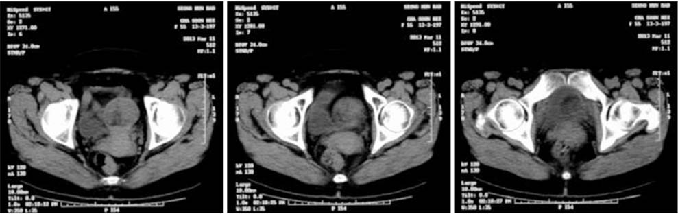
Fig. 1 Abdominal and pelvic computed tomography. Suspicious fat containing pelvic mass, maybe uterine origin.
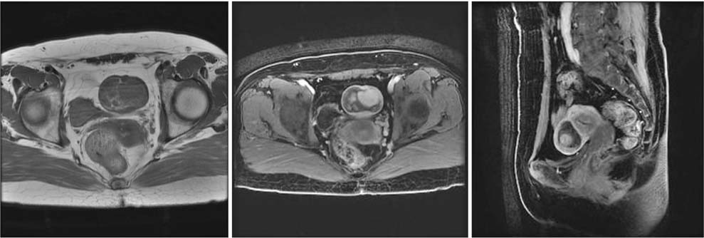
Fig. 2 Pelvic magnetic resonance image. Fat containing heterogeneously enhancing mass, uterus body anterior wall subserosal layer.
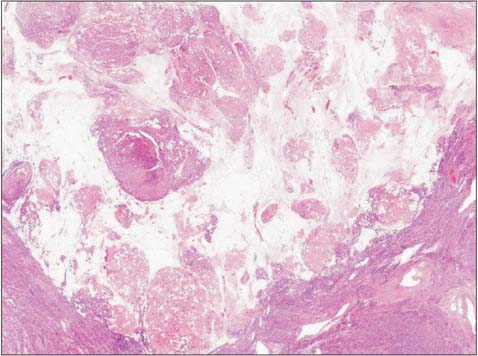
Fig. 3 Admixture of smooth muscle and mature fat cells.
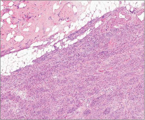
Fig. 4 Highly cellular leiomyoma was composed of interlacing bundles of spindle-shaped smooth muscle cells, no atypia and necrosis (H & E ×40).
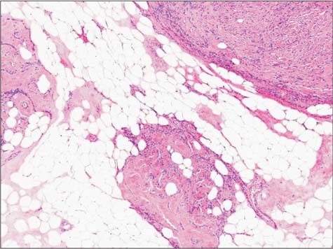
Fig. 5 Mature fat cells (H & E ×100).
Cited by 1 articles
-
Gonadotropin-releasing Hormone Agonist Plus Aromatase Inhibitor in the Treatment of Uterine Leiomyoma in Near Menopause Patient: A Case Series Study
Sanam Moradan
J Menopausal Med. 2018;24(1):62-66. doi: 10.6118/jmm.2018.24.1.62.
Reference
-
1. Manjunatha HK, Ramaswamy AS, Kumar BS, Kumar SP, Krishna L. Lipoleiomyoma of uterus in a postmenopausal woman. J Midlife Health. 2010; 1:86–88.2. Wang X, Kumar D, Seidman JD. Uterine lipoleiomyomas: a clinicopathologic study of 50 cases. Int J Gynecol Pathol. 2006; 25:239–242.3. Aung T, Goto M, Nomoto M, Kitajima S, Douchi T, Yoshinaga M, et al. Uterine lipoleiomyoma: a histopathological review of 17 cases. Pathol Int. 2004; 54:751–758.4. Mira JL. Lipoleiomyoma of the ovary: report of a case and review of the English literature. Int J Gynecol Pathol. 1991; 10:198–202.5. Meis JM, Enzinger FM. Myolipoma of soft tissue. Am J Surg Pathol. 1991; 15:121–125.6. Wahal SP, Mardi K. Lipoleiomyoma of uterus and lipoma of broad ligament--a rare entity. J Cancer Res Ther. 2014; 10:434–436.7. Kumar S, Garg S, Rana P, Hasija S, Kataria SP, Sen R. Lipoleiomyoma of uterus: uncommon incidental finding. Gynecol Obstet. 2013; 3:145.8. Salman MC, Atak Z, Usubutun A, Yuce K. Lipoleiomyoma of broad ligament mimicking ovarian cancer in a postmenopausal patient: case report and literature review. J Gynecol Oncol. 2010; 21:62–64.9. Dalen T, Coebergh JW, Casparie MK, Gimbrére CH, Hoekstra HJ, Van Geel BN, et al. Soft tissue sarcoma: the predominant primary malignancy in the retroperitoneum. Sarcoma. 2001; 5:5–8.10. Erzen D, Sencar M, Novak J. Retroperitoneal sarcoma: 25 years of experience with aggressive surgical treatment at the Institute of Oncology, Ljubljana. J Surg Oncol. 2005; 91:1–9.11. Jeong GA. Retroperitoneal leiomyoma of the uterus mimicking sarcoma in perimenopausal woman: case report. J Menopausal Med. 2014; 20:133–137.12. Lau LU, Thoeni RF. Case report. Uterine lipoma: advantage of MRI over ultrasound. Br J Radiol. 2005; 78:72–74.13. Kim HG, Song YJ, Na YJ, Choi OH. A case of torsion of a subserosal leiomyoma. J Menopausal Med. 2013; 19:147–150.14. Terada T. Giant subserosal lipoleiomyomas of the uterine cervix and corpus: a report of 2 cases. Appl Immunohistochem Mol Morphol. 2015; 23:e1–e3.15. Akbulut M, Soysal ME, Duzcan SE. Giant lipoleiomyoma of the uterine corpus. Arch Gynecol Obstet. 2008; 278:291–293.16. Lin KC, Sheu BC, Huang SC. Lipoleiomyoma of the uterus. Int J Gynaecol Obstet. 1999; 67:47–49.17. Chang IJ, Hong GY, Oh YL, Kim BR, Park SN, Lee HH, et al. Effects of menopausal hormone therapy on uterine myoma in menopausal women. J Menopausal Med. 2013; 19:123–129.18. Lee HP, Tseng HH, Hsieh PP, Shih TF. Uterine lipoleiomyosarcoma: report of 2 cases and review of the literature. Int J Gynecol Pathol. 2012; 31:358–363.19. McDonald AG, Dal Cin P, Ganguly A, Campbell S, Imai Y, Rosenberg AE, et al. Liposarcoma arising in uterine lipoleiomyoma: a report of 3 cases and review of the literature. Am J Surg Pathol. 2011; 35:221–227.20. Vural C, Özen Ö, Demirhan B. Intravenous lipoleiomyomatosis of uterus with cardiac extension: a case report. Pathol Res Pract. 2011; 207:131–134.21. Lee P, Park JH, Yun BH, Lim KJ, Jeon YE, Seo SK, et al. Endometriosis in a postmenopausal woman 16 years after a hysterectomy: a case report. J Korean Soc Menopause. 2011; 17:47–51.
- Full Text Links
-
- Actions
-
Cited
- CITED
-
- Close
- Share
-
- Similar articles
-
- Lipoleiomyoma of broad ligament mimicking ovarian cancer in a postmenopausal patient: case report and literature review
- Exophytic Lipoleiomyoma of the Uterus Mimicking Ovarian Teratoma: A Case Report
- Lipoleiomyoma of the uterus: a case report
- A uterine lipoleiomyoma of the broad ligament mimicking an ovarian tumor
- Extraabdominal parasitic lipoleiomyoma
