A Case of Double Primary Lung Cancer that was Diagnosed by Percutaneous Localization with using a Hook Wire
- Affiliations
-
- 1Department of Internal Medicine, Konyang University College of Medicine, Daejeon, Korea. sk1609@hanmail.net
- 2Department of Diagnostic Radiology, Konyang University College of Medicine, Daejeon, Korea.
- 3Department of Chest Surgery, Konyang University College of Medicine, Daejeon, Korea.
- KMID: 2200034
- DOI: http://doi.org/10.6058/jlc.2008.7.2.93
Abstract
- With the progress of computed tomography (CT), the detection of small pulmonary nodules has been increased. The conventional diagnostic modalities for tissue confirmation, such as bronchoscopic biopsy or transthoracic needle biopsy, may not be successful in some cases. Too small a nodule or the nodules located far from the pleural surface can be marked and localized with device preoperatively and then this tissue can be obtained surgically. CT-guided hook wire fixation is useful in marking pulmonary nodules and there are few complications with this procedure. We report here on a case of double primary lung cancer that was diagnosed by percutaneous localization with using a hook wire
MeSH Terms
Figure
-
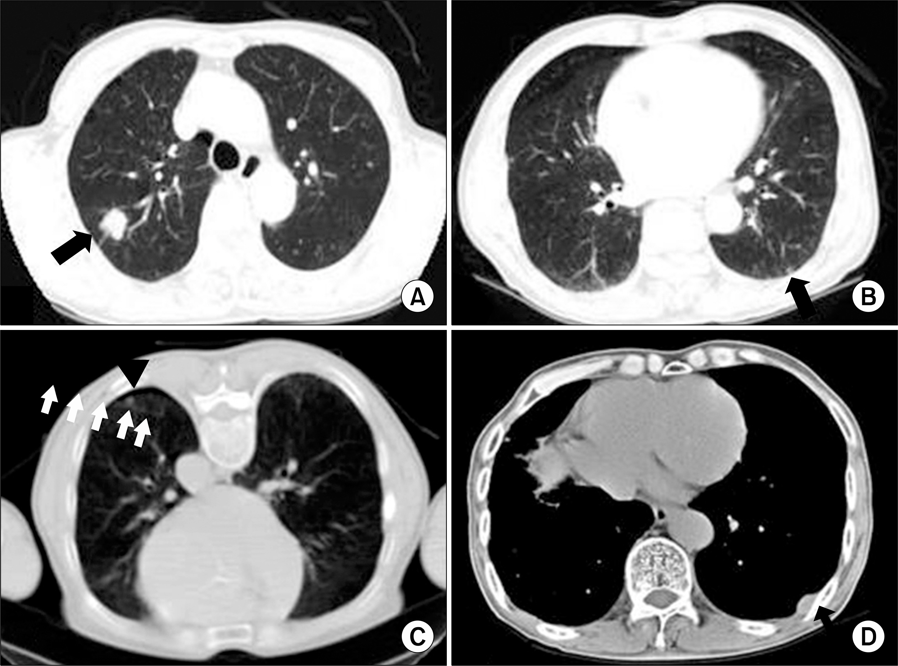
Fig. 1. The initial chest CT scan showed (A) about a 1.7×1.5 cm sized well-defined nodule with a lobulated shape in the right upper lung (solid arrow) and (B) a well-defined small nodule in the left lower lung just beneath the pleura (solid arrow). (C) Before the operation, the small nodule (solid triangle) in the left lower lung was marked and localized with using a hook wire under CT guidance (white arrows). (D) Two years after the operation, the chest CT showed about 2.8 cm sized newly developed heterogeneously enhancing lesion in the left posterior chest wall (solid black arrow).
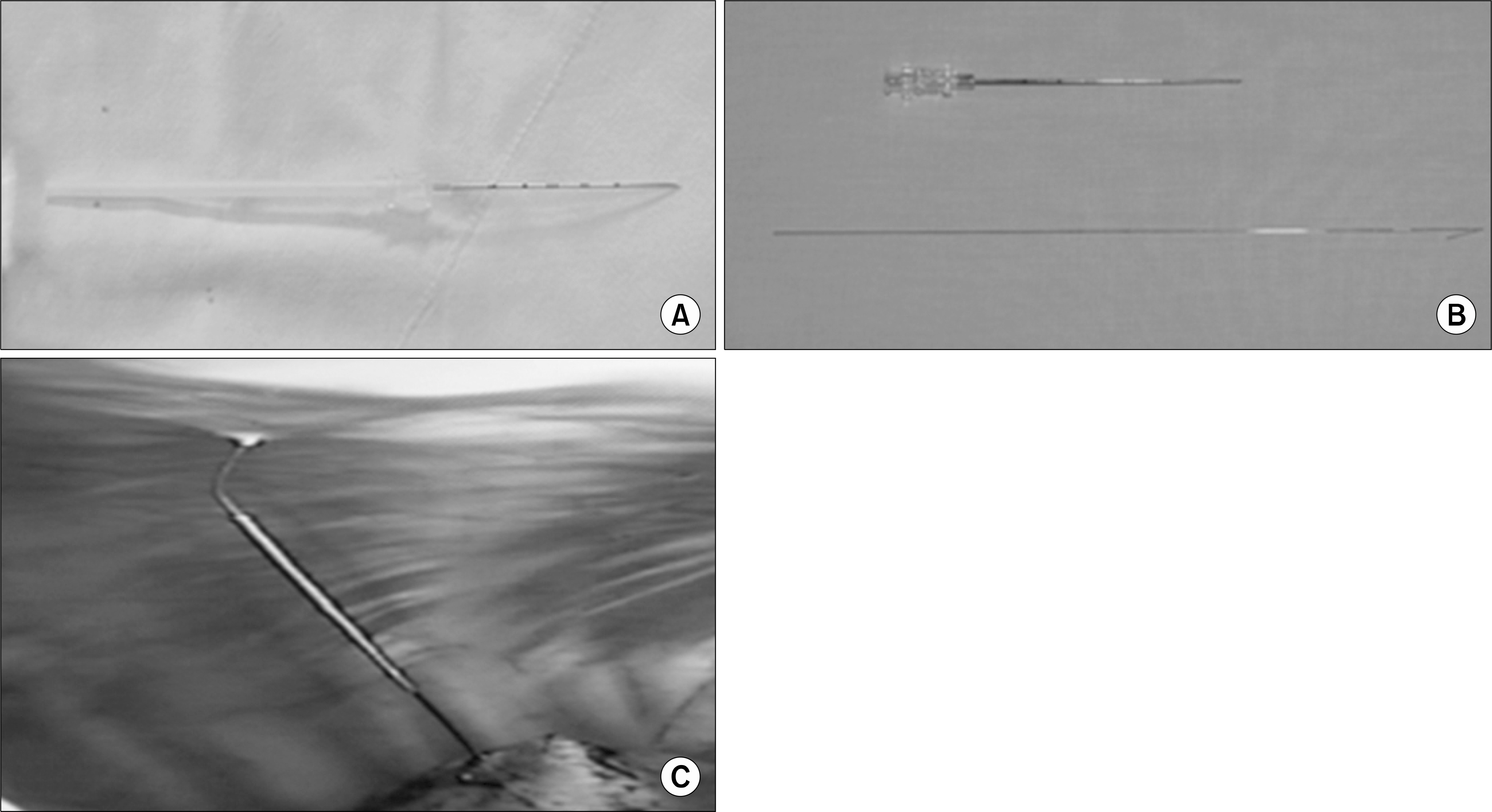
Fig. 2. (A) The hook wire system is composed of a long cannula needle and a hook wire. When the cannula needle tip is placed in a proper position, the hook wire is advanced along the cannula. (B) The 20 gauge, 7.5 cm long cannula needle (top) and the hook wire (bottom). (C) Localization of the small nodule with the hook wire during video-assisted thoracoscopic surgery.
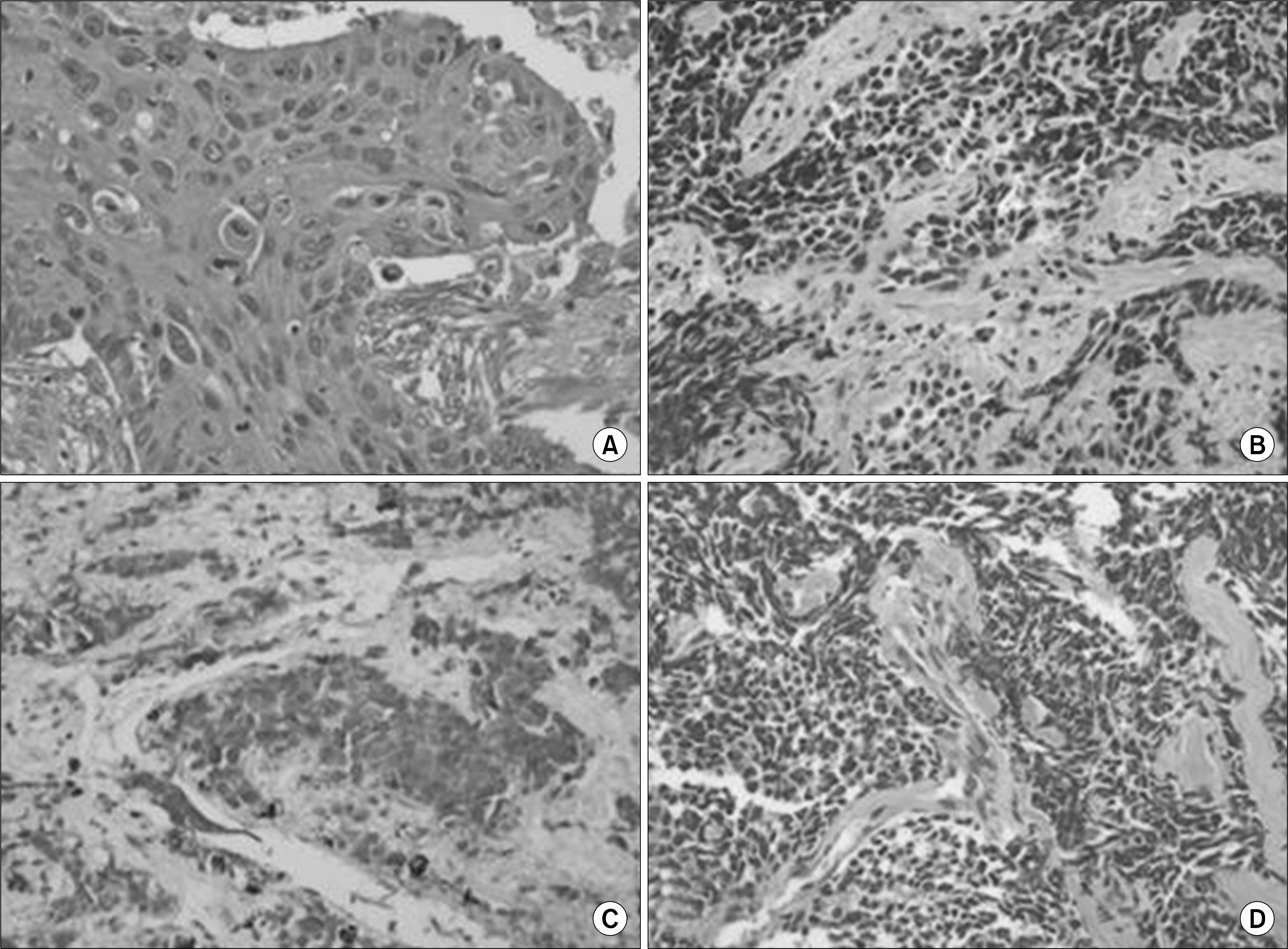
Fig. 3. The report from the pathologist revealed (A) squamous cell carcinoma for the nodule of the right upper lobe (H&E stain, ×200) and (B) small cell carcinoma for the lesion in the left lower lobe, which was obtained by wedge resection after localization with using a hook wire (H&E stain, ×200). (C) The tumor cells for the nodule of left lower lobe were positive for chromogranin, which is an immunohistochemistry stain. (D) Two years after the operation, metastatic small cell carcinoma was shown from the pleural biopsy (H&E stain, ×200).
Reference
-
References
1. Park JK, Sa YJ, Jung JI. Radiologic evaluation for differentiating benign from malignant solitary pulmonary nodule. Korean J Thorac Cardiovasc Surg. 2003; 36:943–951.2. Keogan MT, Tung KT, Kaplan DK, Goldstraw PJ, Hansell DM. The significance of pulmonary nodules detected on CT staging for lung cancer. Clin Radiol. 1993; 48:94–96.
Article3. Lee GI, Lee SS, Won GT, et al. The clinical study of the solitary pulmonary nodule. Korean J Intern Med. 1993; 44:163–170.4. Suzuki K, Nagai K, Yoshida J, et al. Video-assisted thoracoscopic surgery for small indeterminate pulmonary nodules: indications for preoperative marking. Chest. 1999; 115:563–568.5. Saito H, Minamiya Y, Matsuzaki I, et al. Indication for preoperative localization of small peripheral pulmonary nodules in thoracoscopic surgery. J Thorac Cardiovasc Surg. 2002; 124:1198–1202.
Article6. Vandoni RE, Cuttat JF, Wicky S, Suter M. CT-guided methylene-blue labelling before thoracoscopic resection of pulmonary nodules. Eur J Cardiothorac Surg. 1998; 14:265–270.
Article7. Wicky S, Mayor B, Cuttat JF, Schnyder P. CT-guided localizations of pulmonary nodules with methylene blue injections for thoracoscopic resections. Chest. 1994; 106:1326–1328.
Article8. Iwasaki Y, Nagata K, Yuba T, et al. Fluoroscopy-guided barium marking for localizing small pulmonary lesions before video-assisted thoracic surgery. Respir Med. 2005; 99:285–289.
Article9. Tsuchida M, Yamato Y, Aoki T, et al. CT-guided agar marking for localization of nonpalpable peripheral pulmonary lesions. Chest. 1999; 116:139–143.
Article10. Chen YR, Yeow KM, Lee JY, et al. CT-guided hook wire localization of subpleural lung lesions for video-assisted thoracoscopic surgery (VATS). J Formos Med Assoc. 2007; 106:911–918.
Article
- Full Text Links
-
- Actions
-
Cited
- CITED
-
- Close
- Share
-
- Similar articles
-
- Computed Tomography-guided Localization with a Hook-wire Followed by Video-assistedThoracic Surgery for Small Intrapulmonary and Ground Glass Opacity Lesions
- Chest Wall Implantation of Lung Cancer Following CT-Guided Wire Localization: A Case Report
- Comparison of inclination and vertical changes between single-wire and double-wire retraction techniques in lingual orthodontics
- US-Guided Preoperative Hook-Wire Localization of Nonpalpable Breast Lesions
- A Case of Synchronous Double Primary Cancer with Esophageal Small Cell Carcinoma and Lung Squamous Cell Carcinoma
