J Lung Cancer.
2010 Jun;9(1):24-25. 10.6058/jlc.2010.9.1.24.
Small Cell Lung Cancer at Subcarina Presenting as a Metastatic Brain Tumor
- Affiliations
-
- 1Department of Internal Medicine, Bundang CHA Hospital, CHA University College of Medicine, Seongnam, Korea. jhcmd@hanmail.net
- KMID: 2200026
- DOI: http://doi.org/10.6058/jlc.2010.9.1.24
Abstract
- A 59-year-old man was rushed to the emergency room. The patient complained of headache with impaired memory function. Brain MRI showed a necrotic tumor in Lt cerebral hemisphere, with severe peritumoral edema (Fig. 1). Pathologic examination of the brain lesion confirmed that the tumor was a small cell lung cancer (SCLC). Chest computed tomography revealed a large soft tissue mass with central necrosis at subcarinal area in spite of an initial normal chest X-ray (Fig. 2). Bronchoscopic biopsy of the polypoid mass at subcarina revealed that the mass was a SCLC (Fig. 3). This is the case of SCLC only with an extrapulmonary symptoms despite of a normal chest X-ray. When metastatic brain tumor was found, appropriate chest evaluation should be performed even though chest X-ray was normal because brain is a common site of invasion of lung cancer.
MeSH Terms
Figure
-

Fig. 1. Initial Brain MRI shows necrotic tumors in Lt cerebral hemisphere, with severe peritumoral edema.
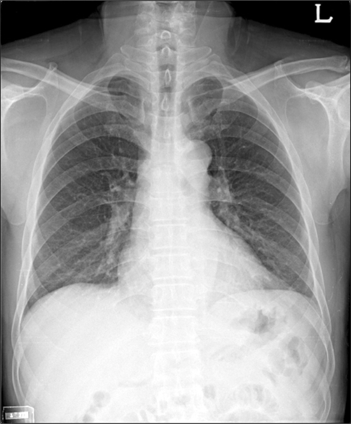
Fig. 2. Initial chest PA shows no active lung lesion.
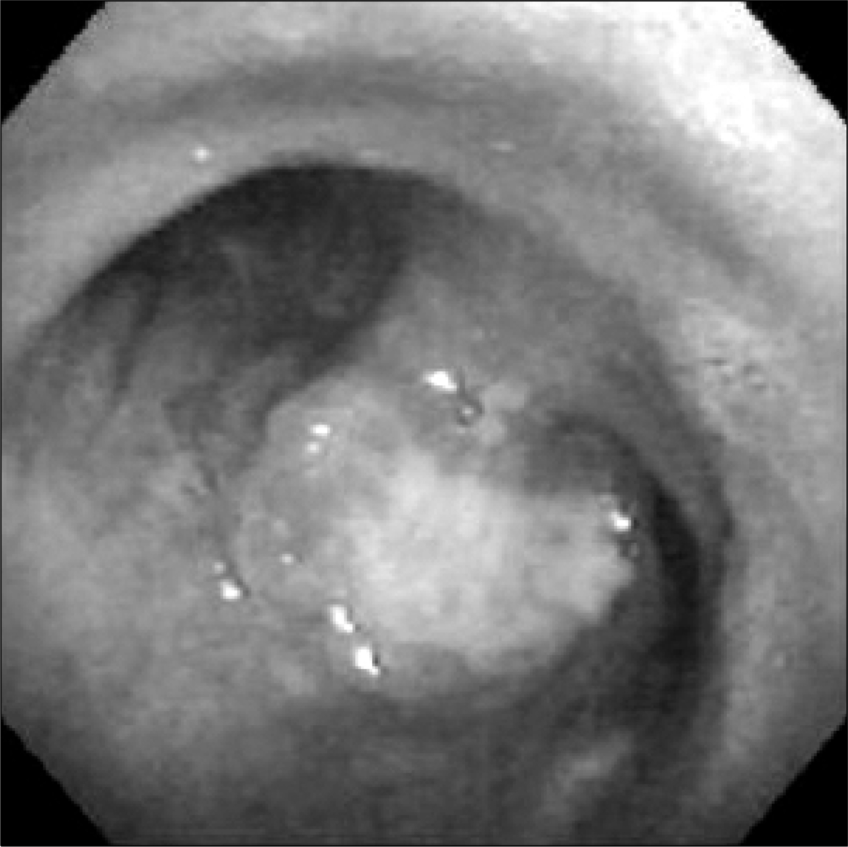
Fig. 3. Bronchoscopy shows the polypoid mass at subcarina.
- Full Text Links
-
- Actions
-
Cited
- CITED
-
- Close
- Share
-
- Similar articles
-
- A Case of Metastatic Gastric Cancer Resulting from Small Cell Lung Cancer
- Radiotherapy of Brain Metastases from Lung Cancer
- Two Cases of Cutaneous Metastasis from Small Cell Lung Cancer
- A Case of Iris Metastasis from Non-small Cell Lung Cancer
- An Unusual Case of Metastatic Non-Small Cell Lung Cancer Misidentified as Anaplastic Thyroid Cancer
