Clinical case of implant restoration using customized healing abutment
- Affiliations
-
- 1Department of Prosthodontics, College of Dentistry and Advanced Dental Device Development Institute, Kyungpook National University, Daegu, Republic of Korea. kblee@knu.ac.kr
- KMID: 2195252
- DOI: http://doi.org/10.4047/jkap.2015.53.3.222
Abstract
- Aesthetic impression is emphasized in the recent field of implant restoration. However, there is limitation of creating proper shape of soft tissue as well as cervical emergence profile due to the use of pre-existing healing abutment in the process of initial post-operative soft tissue healing period. Designing the shape of abutment into the final customized abutment instead of its original shape helped to achieve more aesthetic implant restoration by applying healing abutment which could minimize the malposition and recession of soft tissue. In this study, soft tissue healing was promoted using the post-operative customized healing abutment and thereby obtained the result of more aesthetic and functional restoration by minimizing displacement of soft tissue in the process of applying final customized abutment.
Figure
-

Fig. 1. Panoramic radiograph on first visit.

Fig. 2. Postoperative intraoral view. (A) Frontal view, (B, C) Occlusal view, (D, E) Buccal view.
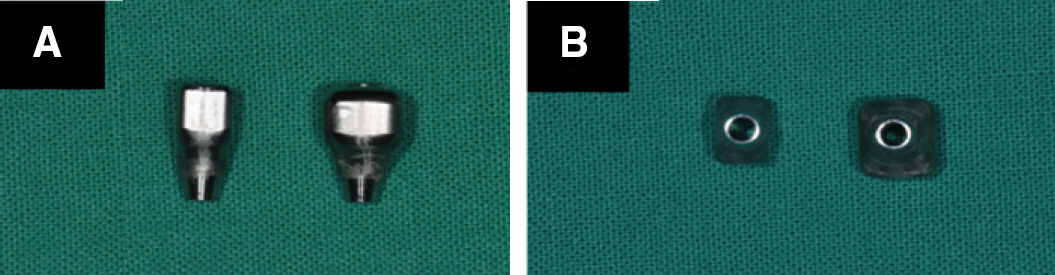
Fig. 3. Customized healing abutment used in this study. (A) Lateral view of Lt. 2 nd premolar, 1st molar. (B) bottom view of Lt. Mx. 2nd premolar, 1st molar (Raphabio Co., Deagu, Korea).
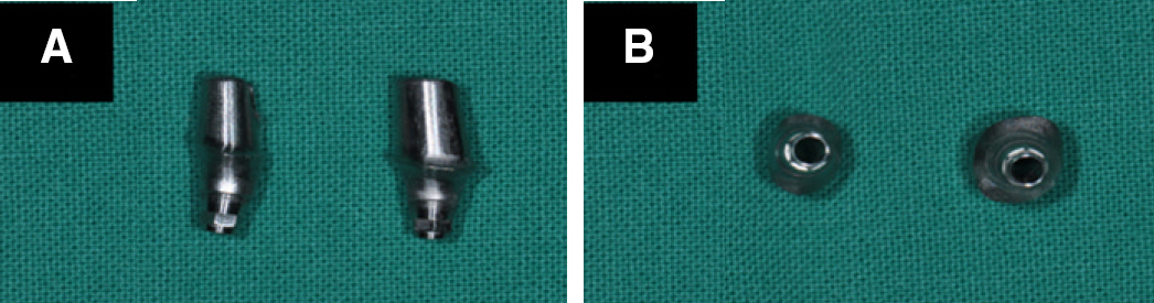
Fig. 4. Customized abutment used in this study. (A) Lateral view of Lt. 2 nd premolar, 1 st molar. (B) bottom view of Lt. Mx. 2 nd premolar, 1 st molar (Myplant, Raphabio Co., Deagu, Korea).
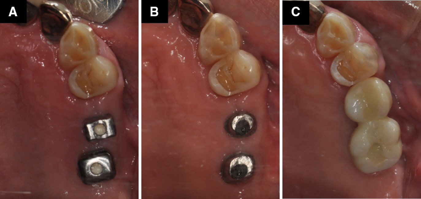
Fig. 5. Clinical photographs of this case. (A) Intra oral photographs of customized healing abutment after surgery (B) Customized abutment connected for final prosthesis (C) final prosthesis.
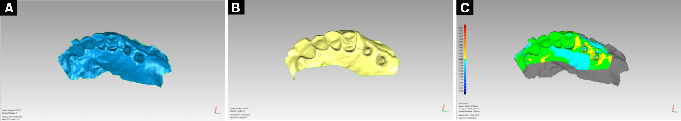
Fig. 6. Scanned data was used intraoal scanner. (A) virtual model is scanned after customized healing abutment was removed. (B) virtual model is re-scanned after final abutment was removed. (C) Superimposed images of two scan images.

Fig. 7. Analysis of difference between the two virtual models (Customized healing abutment used-dotted line / final abutment used-purple line). (A) Showed line to cross section on the superimposed images (B) Cross-sectional view of the buccolingual on Lt. Mx. 2nd premolar (C) Cross-sectional view of the buccolingual on Lt. Mx. 1st molar (D) Cross-sectional views of the mesiodistal Lt. Mx. 2nd premolar and Lt. Mx. 1st molar.
Reference
-
1. Phillips K, Kois JC. Aesthetic peri-implant site development. The restorative connection. Dent Clin North Am. 1998; 42:57–70.2. Davarpanah M, Martinez H, Tecucianu JF. Apical-coronal implant position: recent surgical proposals. Technical note. Int J Oral Maxillofac Implants. 2000; 15:865–72.3. Funato A, Salama MA, Ishikawa T, Garber DA, Salama H. Timing, positioning, and sequential staging in esthetic implant therapy: a four-dimensional perspective. Int J Periodontics Restorative Dent. 2007; 27:313–23.4. Garber DA, Belser UC. Restoration-driven implant placement with restoration-generated site development. Compend Contin Educ Dent. 1995; 16(796):798–802. 804.5. Grunder U, Gracis S, Capelli M. Influence of the 3-D bone-to-implant relationship on esthetics. Int J Periodontics Restorative Dent. 2005; 25:113–9.6. Kois JC. Predictable single tooth peri-implant esthetics: five diagnostic keys. Compend Contin Educ Dent. 2001; 22:199–206.7. Smukler H, Castellucci F, Capri D. The role of the implant housing in obtaining aesthetics: Part 2. Customizing the peri-implant soft tissue. Pract Proced Aesthet Dent. 2003; 15:487–90.8. Weisgold AS, Arnoux JP, Lu J. Single-tooth anterior implant: a world of caution. Part I. J Esthet Dent. 1997; 9:225–33.9. Smukler H, Castellucci F, Capri D. The role of the implant housing in obtaining aesthetics: generation of peri-implant gin-givae and papillae-Part 1. Pract Proced Aesthet Dent. 2003; 15:141–9.10. Bichacho N, Landsberg CJ. Single implant restorations: prosthetically induced soft tissue topography. Pract Periodontics Aesthet Dent. 1997; 9:745–52.11. Cooper LF. Objective criteria: guiding and evaluating dental implant esthetics. J Esthet Restor Dent. 2008; 20:195–205.
Article12. Su H, Gonzalez-Martin O, Weisgold A, Lee E. Considerations of implant abutment and crown contour: critical contour and sub-critical contour. Int J Periodontics Restorative Dent. 2010; 30:335–43.
- Full Text Links
-
- Actions
-
Cited
- CITED
-
- Close
- Share
-
- Similar articles
-
- Improvement of peri-implant complications through customized prosthesis restoration allowing soft tissue space: a case report
- SINGLE TOOTH IMPLANT RESTORATION USING COMBINATION IMPLANT CROWN : A CASE REPORT
- Maxillary anterior single implant prosthesis
- Posterior single implant prosthesis using scannable healing abutment
- Restoration of implant-supported fixed dental prosthesis using the automatic abutment superimposition function of the intraoral scanner in partially edentulous patients
