J Korean Neurosurg Soc.
2016 May;59(3):310-313. 10.3340/jkns.2016.59.3.310.
Posttraumatic Intracranial Tuberculous Subdural Empyema in a Patient with Skull Fracture
- Affiliations
-
- 1Department of Neurosurgery, Kangwon National University Hospital, Kangwon National University School of Medicine, Chuncheon, Korea. rabbit3540@empas.com
- 2Department of Pathology, Kangwon National University Hospital, Kangwon National University School of Medicine, Chuncheon, Korea.
- KMID: 2192113
- DOI: http://doi.org/10.3340/jkns.2016.59.3.310
Abstract
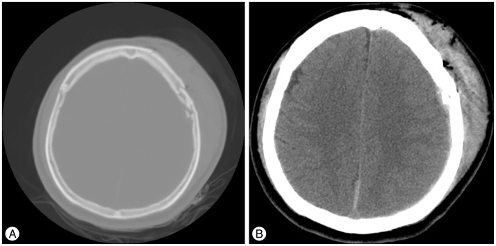
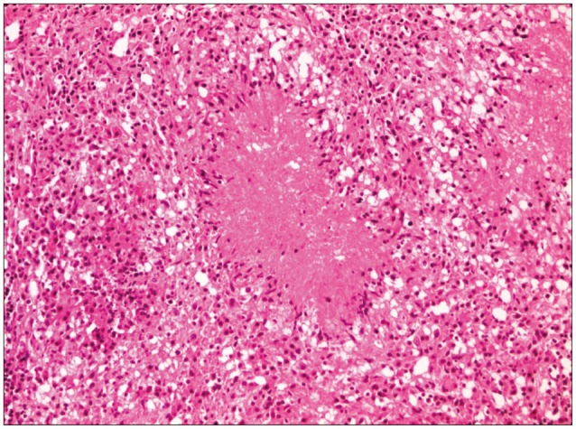
- Intracranial tuberculous subdural empyema (ITSE) is extremely rare. To our knowledge, only four cases of microbiologically confirmed ITSE have been reported in the English literature to date. Most cases have arisen in patients with pulmonary tuberculosis regardless of trauma. A 46-year-old man presented to the emergency department after a fall. On arrival, he complained of pain in his head, face, chest and left arm. He was alert and oriented. An initial neurological examination was normal. Radiologic evaluation revealed multiple fractures of his skull, ribs, left scapula and radius. Though he had suffered extensive skull fractures of his cranium, maxilla, zygoma and orbital wall, the sustained cerebral contusion and hemorrhage were mild. Eighteen days later, he suddenly experienced a general tonic-clonic seizure. Radiologic evaluation revealed a subdural empyema in the left occipital area that was not present on admission. We performed a craniotomy, and the empyema was completely removed. Microbiological examination identified Mycobacterium tuberculosis (M. tuberculosis). After eighteen months of anti-tuberculous treatment, the empyema disappeared completely. This case demonstrates that tuberculosis can induce empyema in patients with skull fractures. Thus, we recommend that M. tuberculosis should be considered as the probable pathogen in cases with posttraumatic empyema.
Keyword
MeSH Terms
Figure
Reference
-
1. Ansari MK, Jha S. Tuberculous brain abscess in an immunocompetent adolescent. J Nat Sci Biol Med. 2014; 5:170–172. PMID: 24678219.
Article2. Banerjee AD, Pandey P, Ambekar S, Chandramouli BA. Pediatric intracranial subdural empyema caused by Mycobacterium tuberculosis--a case report and review of literature. Childs Nerv Syst. 2010; 26:1117–1120. PMID: 20437243.
Article3. Cayli SR, Onal C, Koçak A, Onmuş SH, Tekiner A. An unusual presentation of neurotuberculosis: subdural empyema. Case report. J Neurosurg. 2001; 94:988–991. PMID: 11409530.4. Eftekhar B, Ghodsi M, Nejat F, Ketabchi E, Esmaeeli B. Prophylactic administration of ceftriaxone for the prevention of meningitis after traumatic pneumocephalus: results of a clinical trial. J Neurosurg. 2004; 101:757–761. PMID: 15540912.
Article5. Katti MK. Pathogenesis, diagnosis, treatment, and outcome aspects of cerebral tuberculosis. Med Sci Monit. 2004; 10:RA215–RA229. PMID: 15328498.6. Lee SK, Kim SW. Fatal subdural empyema following pyogenic meningitis. J Korean Neurosurg Soc. 2011; 49:175–177. PMID: 21556239.
Article7. Madhavan K, Widi G, Shah A, Petito C, Gallo BV, Komotar RJ. Tuberculoma of the brain with unknown primary infection in an immunocompetent host. J Clin Neurosci. 2012; 19:1320–1322. PMID: 22727748.
Article8. Scholsem M, Scholtes F, Collignon F, Robe P, Dubuisson A, Kaschten B, et al. Surgical management of anterior cranial base fractures with cerebrospinal fluid fistulae: a single-institution experience. Neurosurgery. 2008; 62:463–439. discussion 469-471. PMID: 18382325.9. Servais L, Fonteyne C, Christophe C, Prudhon V, Brihaye P, Biarent D, et al. Meningitis following basal skull fracture in two in-line skaters. Childs Nerv Syst. 2005; 21:339–342. PMID: 15798922.
Article10. Sonig A, Thakur JD, Chittiboina P, Khan IS, Nanda A. Is posttraumatic cerebrospinal fluid fistula a predictor of posttraumatic meningitis? A US Nationwide Inpatient Sample database study. Neurosurg Focus. 2012; 32:E4. PMID: 22655693.
Article11. Turel MK, Moorthy RK, Rajshekhar V. Multidrug-resistant tuberculous subdural empyema with secondary methicillin-resistant Staphylococcus aureus infection: an unusual presentation of cranial tuberculosis in an infant. Neurol India. 2012; 60:231–234. PMID: 22626710.
Article


