J Korean Neurosurg Soc.
2016 Mar;59(2):165-167. 10.3340/jkns.2016.59.2.165.
Case of Langerhans Cell Histiocytosis That Mimics Meningioma in CT and MRI
- Affiliations
-
- 1Department of Medical Imaging, Jinan Military General Hospital, Jinan, China. cjr.sungang@vip.163.com
- 2Department of Radiology, The Branch of Taian Central Hospital, Taian, China.
- KMID: 2192049
- DOI: http://doi.org/10.3340/jkns.2016.59.2.165
Abstract
- Langerhans cell histiocytosis (LCH) is a rare disorder histologically characterized by the proliferation of Langerhans cells. Here we present the case of a 13-year-old girl with LCH wherein CT and MRI results led us to an initially incorrect diagnosis of meningioma. The diagnosis was corrected to LCH based on pathology findings. An intracranial mass was found mainly in the dura mater, with thickening of the surrounding dura. It appeared to be growing downward from the calvaria, pressing on underlying brain tissue, and had infiltrated the inner skull, causing a bone defect. The lesion was calcified with the typical dural tail sign. The dural origin of the lesion was verified upon surgical dissection. There are no previous reports in the literature describing LCH of dural origin presenting in young patients with typical dural tail signs and meningioma-like imaging findings. The current case report underscores the need for thorough histological and immunocytochemical examinations in LCH differential diagnosis.
Keyword
MeSH Terms
Figure
-
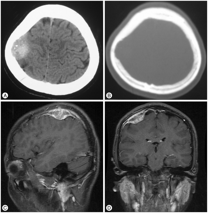
Fig. 1 A : Plain CT scan. Soft-tissue window, high-density mass on the right calvaria with internal calcification spots. B : Plain CT scan. Bone window and adjacent bone deficit with unclear edges. C and D : Enhanced MRI revealing an irregularly shaped tumor with clear boundaries and a wide base attached to the meninges (sagittal and coronal planes, respectively).
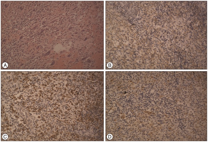
Fig. 2 Histopathological demonstration of LCH. Hematoxylin-eosin staining of the resected mass revealed a large number of Langerhans cells, multinucleated giant cells, and diffuse eosinophil and lymphocyte infiltration (A). Immunohistochemistry revealed the presence of membranous CD1a (B), cytoplasmic CD68 (C), and nuclear S-100 (D). Original magnification : ×200.
Reference
-
1. Anagnostou E, Papageorgiou SG, Potagas C, Alexakis T, Kalfakis N, Anastasopoulos D. Square-wave jerks and smooth pursuit impairment as subtle early signs of brain involvement in Langerhans' cell histiocytosis. Clin Neurol Neurosurg. 2008; 110:286–290. PMID: 18078708.
Article2. Aricò M. Langerhans cell histiocytosis in adults : more questions than answers? Eur J Cancer. 2004; 40:1467–1473. PMID: 15196529.3. Baba T, Ibayashi Y, Morimoto S, Niwa J, Tanabe S, Hashi K. [A case of dural type of histiocytosis X presenting as a mass lesion in the tentorium cerebelli]. No Shinkei Geka. 1994; 22:471–476. PMID: 8196835.4. D'Ambrosio N, Soohoo S, Warshall C, Johnson A, Karimi S. Craniofacial and intracranial manifestations of langerhans cell histiocytosis : report of findings in 100 patients. AJR Am J Roentgenol. 2008; 191:589–597. PMID: 18647937.5. Holbrook TJ, Fogo A, Smith HP. Histiocytosis X involving the cervical dura. Childs Nerv Syst. 1987; 3:50–52. PMID: 3496150.
Article6. Kasper EM, Aguirre-Padilla DH, Alter RY, Anderson M. Histiocytosis X : Characteristics, behavior, and treatments as illustrated in a case series. Surg Neurol Int. 2011; 2:57. PMID: 21697965.
Article7. Louis D, Ohgaki H, Wiestler OD, Cavenee WK, Burger PC, Jouvet A, et al. The 2007 WHO Classification of Tumours of the Central Nervous System. Acta Neuropathol. 2007; 114:97–109. PMID: 17618441.
Article8. Mosiewicz A, Rola R, Jarosz B, Trojanowska A, Trojanowski T. Langerhans cell histiocytosis of the parietal bone with epidural and extracranial expansion - case report and a review of the literature. Neurol Neurochir Pol. 2010; 44:196–203. PMID: 20496290.
Article9. Prayer D, Grois N, Prosch H, Gadner H, Barkovich AJ. MR imaging presentation of intracranial disease associated with Langerhans cell histiocytosis. AJNR Am J Neuroradiol. 2004; 25:880–891. PMID: 15140741.
- Full Text Links
-
- Actions
-
Cited
- CITED
-
- Close
- Share
-
- Similar articles
-
- A Case of Pulmonary Langerhans Cell Histiocytosis with Pneumothorax
- A Case of Langerhans Cell Histiocytosis Mimicking Periorbital Cellulitis
- Spontaneous Pneumothorax due to Pulmonary Invasion in Multisystemic Langerhans Cell Histiocytosis: A case report
- A Case of Gastric Langerhans Cell Histiocytosis Showing Hypertrophic Folds
- A Case of Orbital Langerhans' cell histiocytosis
