Rupturing Anterior Communicating Artery Aneurysm during Computed Tomography Angiography: Three-Dimensional Visualization of Bleeding into the Septum Pellucidum and the Lateral Ventricle
- Affiliations
-
- 1Department of Neurosurgery, Dongsan Medical Center, Keimyung University School of Medicine, Daegu, Korea. bach1158@dsmc.or.kr
- KMID: 2191117
- DOI: http://doi.org/10.3340/jkns.2014.55.6.357
Abstract
- Computed tomography angiography (CTA) is commonly used in setting of subarachnoid hemorrhage, but imaging features of aneurysm rupturing taking place at the time of scanning has rarely been described. The author reports a case of actively rebleeding aneurysm of the anterior communicating artery with intraventricular extravasation on the hyperacute CTA imaging. The rebleeding route, not into the third ventricle but into the lateral ventricles, can be visualized by real-time three-dimensional CT pictures. The hemorrhage broke the septum pellucidum and the lamina rostralis rather than the lamina terminalis.
Keyword
MeSH Terms
Figure
-

Fig. 1 A : Initial plain CT scan shows thin SAH and subdural hematoma. B : There is no cavum septum pellucidum on the vertex cut. C : Source image of CTA reveals saccular aneurysms at the anterior communicating artery (arrow) and the left middle cerebral artery. D and E : Delayed images immediately after CTA show extensive SAH associated with intracerebral and intraventricular hemorrhages as well as blood within the septum pellucidum. CTA : computed tomography angiography, SAH : subarachnoid hemorrhage.
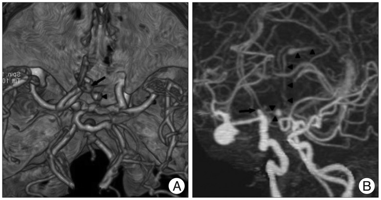
Fig. 2 A : Three-dimensional reconstruction of CTA depicts the spatial relationship between the aneurysm (arrow) and extravasated contrast medium (arrowhead). B : CTA with maximum intensity projection outlines the entire region of extravasated fresh blood (arrowheads) spouting from the ruptured ACoA aneurysm (arrow). ACoA : anterior communicating artery, CTA : computed tomography angiography.
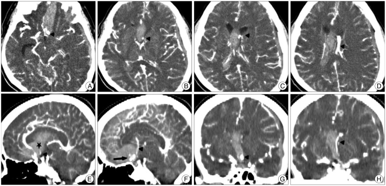
Fig. 3 A-D : Axial images of CTA demonstrate the caudorostral pathway of the extravasated contrast medium (arrowheads) into the preexisting hematoma of lower density from the interhemispheric cistern into the septum pellucidum and into the frontal horn and body of the lateral ventricle. E and F : Sagittal CTA views illustrate the diffusion of the contrast medium (arrowheads) from the rebleeding ACoA aneurysm (arrow) through the lamina rostralis (asterisk) into the septum pellucidum without penetrating into the lamina terminalis. G and H : Coronal reformatted CT scan clearly shows the ejected blood (arrowheads) which pass through the septum pellucidum up to the frontal horn of the left lateral ventricle. ACoA : anterior communicating artery, CTA : computed tomography angiography.

Fig. 4 Coronal T2-weighted magnetic resonance image shows the thin strip of white matter of the lamina rostralis (arrowhead). This separates the anterior interhemispheric cistern (lower arrow) from the space between the leaves of the septum pellucidum (upper arrow) as well as the floor of the frontal horns in the mature brain.
Reference
-
1. Dehdashti AR, Rufenacht DA, Delavelle J, Reverdin A, de Tribolet N. Therapeutic decision and management of aneurysmal subarachnoid haemorrhage based on computed tomographic angiography. Br J Neurosurg. 2003; 17:46–53. PMID: 12779201.
Article2. Desai S, Friedman JA, Hlavin J, Kash F. Actively bleeding intracranial aneurysm demonstrated by CT angiography. Clin Neurol Neurosurg. 2009; 111:94–96. PMID: 18980796.
Article3. Fujii Y, Takeuchi S, Sasaki O, Minakawa T, Koike T, Tanaka R. Ultra-early rebleeding in spontaneous subarachnoid hemorrhage. J Neurosurg. 1996; 84:35–42. PMID: 8613833.
Article4. Gosselin MV, Vieco PT. Active hemorrhage of intracranial aneurysms : diagnosis by CT angiography. J Comput Assist Tomogr. 1997; 21:22–24. PMID: 9022763.
Article5. Hashiguchi A, Mimata C, Ichimura H, Morioka M, Kuratsu J. Rebleeding of ruptured cerebral aneurysms during three-dimensional computed tomographic angiography : report of two cases and literature review. Neurosurg Rev. 2007; 30:151–154. PMID: 17345112.
Article6. Holodny AI, Farkas J, Schlenk R, Maniker A. Demonstration of an actively bleeding aneurysm by CT angiography. AJNR Am J Neuroradiol. 2003; 24:962–964. PMID: 12748102.7. Im SH, Oh CW, Hong SK, Kwon OK, Kim SH. CT angiography demonstration of the development of intraventricular hemorrhage during aneurysm rupture. Clin Neurol Neurosurg. 2007; 109:299–301. PMID: 17207925.
Article8. Kathuria S, Deveikis JP, Westesson PL, Gandhi D. Improved diagnosis of actively bleeding aneurysm on CT angiography using delayed CT images. Eur J Radiol. 2011; 79:328–331. PMID: 20227214.
Article9. Kier EL, Truwit CL. The lamina rostralis : modification of concepts concerning the anatomy, embryology, and MR appearance of the rostrum of the corpus callosum. AJNR Am J Neuroradiol. 1997; 18:715–722. PMID: 9127036.10. Klisch J, Weyerbrock A, Spetzger U, Schumacher M. Active bleeding from ruptured cerebral aneurysms during diagnostic angiography: emergency treatment. AJNR Am J Neuroradiol. 2003; 24:2062–2065. PMID: 14625234.11. Kuhn J, Vehlen C, Mennel HD, Mahkorn D, Bewermeyer H. Rupture of an internal carotid artery aneurysm during angiography with leakage of contrast medium via an external ventricular drain. Neuroradiology. 2003; 45:905–907. PMID: 14534767.
Article12. Kusumi M, Yamada M, Kitahara T, Endo M, Kan S, Iida H, et al. Rerupture of cerebral aneurysms during angiography--a retrospective study of 13 patients with subarachnoid hemorrhage. Acta Neurochir (Wien). 2005; 147:831–837. PMID: 15900400.
Article13. Larsen CC, Astrup J. Rebleeding after aneurysmal subarachnoid hemorrhage : a literature review. World Neurosurg. 2013; 79:307–312. PMID: 22722033.
Article14. Nagai M, Koizumi Y, Tsukue J, Watanabe E. A case of extravasation from a cerebral aneurysm during 3-dimensional computed tomography angiography. Surg Neurol. 2008; 69:411–413. PMID: 18261775.
Article15. Naidech AM, Janjua N, Kreiter KT, Ostapkovich ND, Fitzsimmons BF, Parra A, et al. Predictors and impact of aneurysm rebleeding after subarachnoid hemorrhage. Arch Neurol. 2005; 62:410–416. PMID: 15767506.
Article16. Nakatsuka M, Mizuno S, Uchida A. Extravasation on three-dimensional CT angiography in patients with acute subarachnoid hemorrhage and ruptured aneurysm. Neuroradiology. 2002; 44:25–30. PMID: 11942496.
Article17. Ohkuma H, Tsurutani H, Suzuki S. Incidence and significance of early aneurysmal rebleeding before neurosurgical or neurological management. Stroke. 2001; 32:1176–1180. PMID: 11340229.
Article18. Pérez-Núñez A, Alén JF, Ramos A, Millán JM. Aneurysm re-rupture during computed tomography angiography. Acta Radiol. 2006; 47:419–421. PMID: 16739704.
Article19. Saitoh H, Hayakawa K, Nishimura K, Okuno Y, Murayama C, Miyazawa T, et al. Intracarotid blood pressure changes during contrast medium injection. AJNR Am J Neuroradiol. 1996; 17:51–54. PMID: 8770249.20. Scholtes F, Signorelli F, Bojanowski MW. Rupture of anterior communicating artery aneurysms during computed tomography angiography : description of the pathway for intraseptal and intraventricular hemorrhage. J Neurosurg. 2011; 115:617–620. PMID: 21599449.
Article21. Sorimachi T, Takeuchi S, Koike T, Minakawa T, Tanaka R. Intra-aneurysmal pressure changes during angiography in coil embolization. Surg Neurol. 1997; 48:451–457. PMID: 9352808.
Article22. Starke RM, Kim GH, Fernandez A, Komotar RJ, Hickman ZL, Otten ML, et al. Impact of a protocol for acute antifibrinolytic therapy on aneurysm rebleeding after subarachnoid hemorrhage. Stroke. 2008; 39:2617–2621. PMID: 18658042.
Article23. Tsuang FY, Su IC, Chen JY, Lee JE, Lai DM, Tu YK, et al. Hyperacute cerebral aneurysm rerupture during CT angiography. J Neurosurg. 2012; 116:1244–1250. PMID: 22443505.
Article24. Westerlaan HE, van Dijk JM, Jansen-van der Weide MC, de Groot JC, Groen RJ, Mooij JJ, et al. Intracranial aneurysms in patients with subarachnoid hemorrhage : CT angiography as a primary examination tool for diagnosis--systematic review and meta-analysis. Radiology. 2011; 258:134–145. PMID: 20935079.
Article25. Wu TC, Tsui YK, Chen TY, Lin CJ, Wu TC, Tzeng WS. Rebleeding of aneurysmal subarachnoid hemorrhage in computed tomography angiography : risk factor, rebleeding pattern, and outcome analysis. J Comput Assist Tomogr. 2012; 36:103–108. PMID: 22261779.
Article26. Yanaka K, Meguro K, Narushima K, Takano S, Doi M, Nose T. Basal perforating artery aneurysm within the cavum septi pellucidi. Case report. J Neurosurg. 1998; 88:601–604. PMID: 9488321.
- Full Text Links
-
- Actions
-
Cited
- CITED
-
- Close
- Share
-
- Similar articles
-
- Determination of main feeding artery with CT findings in cases of ruptured aneurysm of anterior communicating artery
- Prediction of Parent Artery of Anterior Communicating Artery Aneurysm on CT Angiography
- Aneurysms of Proximal(A1) Segment of Anterior Cerebral Artery
- Rupture of Giant Posterior Communicating Artery Aneurysm During Carotid Angiography: A Case Report
- Rupture of De Novo Anterior Communicating Artery Aneurysm 8 Days after the Clipping of Ruptured Middle Cerebral Artery Aneurysm
