Spinal Cord Ependymoma Associated with Neurofibromatosis 1 : Case Report and Review of the Literature
- Affiliations
-
- 1Department of Neurosurgery, The First Affiliated Hospital of Anhui Medical University, Anhui, China. chw001@163.com
- 2Department of Pathology, The First Affiliated Hospital of Anhui Medical University, Anhui, China.
- KMID: 2191040
- DOI: http://doi.org/10.3340/jkns.2014.55.1.43
Abstract
- Patients with neurofibromatosis 1 (NF1) are predisposed to develop central nervous system tumors, due to the loss of neurofibromin, an inactivator of proto-oncogene Ras. However, to our knowledge, only three cases of ependymomas with NF1 have been reported in the literature. The authors present a case of NF1 patient with a spinal cord ependymoma. She was referred for about half a year history of increasing numbness that progressed from her fingers to her entire body above the bellybutton. Magnetic resonance imaging revealed a relative-demarcated, heterogeneously enhanced mass lesion accompanied by perifocal edema in C5-7 level, a left-sided T11 spinous process heterogeneously enhanced mass in soft tissue, intervertebral disk hernia in L2-5 level, and widespread punctum enhancing lesion in her scalp and in T11-L5 level. The patient underwent C5-7 laminectomies and total excision of the tumor under operative microscope, and intraoperative ultrasonography and physiological monitoring were used during the surgery. Histopathologically, her tumor was found to be a ependymoma without malignant features (grade II in the World Health Organization classification). Therefore, no adjuvant therapy was applied. Following the operation, the patient showed an uneventful clinical recovery with no evidence of tumor recurrence after one year of follow-up.
Keyword
MeSH Terms
-
Central Nervous System Neoplasms
Edema
Ependymoma*
Fingers
Follow-Up Studies
Hernia
Humans
Hypesthesia
Intervertebral Disc
Laminectomy
Magnetic Resonance Imaging
Monitoring, Physiologic
Neurofibromatoses*
Neurofibromatosis 1*
Neurofibromin 1
Proto-Oncogenes
Recurrence
Scalp
Spinal Cord*
Ultrasonography
World Health Organization
Neurofibromin 1
Figure
-
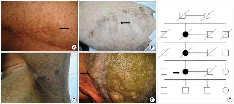
Fig. 1 A : Operative scar on her back and café-au-lait spot (arrow). B : Operative scar on her left thigh and widespread café-au-lait spots (arrow). C : Widespread frecklings axillary. D : Cutaneous neurofibromas and plexiform neurofibroma (arrow). E : Patient's pedigree. The circles represent females and squares represent males. Black represents affected individuals. Oblique line represents dead individuals. The patient is signed in arrow, her mother and maternal grandmother have similar manifestations of neurofibromatosis 1.

Fig. 2 A : Spinal MRI shows a relative-demarcated, heterogeneously enhanced mass lesion accompanied by perifocal edema in C5-7 level (arrow). B : A left-sided T11 spinous process heterogeneously enhanced mass in soft tissue (thick arrow), and intervertebral disk hernia in L2-5 level (thin arrows). C, E, F, and G : Post-gadolinium T1 weighted sagittal imaging revealed widespread punctum enhancing lesion in her scalp and in T11-L5 level. D : Follow-up MRI revealed stable postsurgical changes in the C4-7 level with no evidence of tumor recurrence one year later. H, I, and J : Head MRI suggests no evidences of optic pathway glioma and neurofibromatosis 2.

Fig. 3 A : A yellowish mass with abundance blood supply in the spinal cord is exposed. B : The spinal cord is slit and the tumor was gross-totally resected safely under an operative microscope.
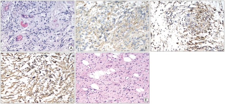
Fig. 4 A : In the H&E staining, the cyst wall consists of glial cells characterized by round, irregular nucei and eosinophilic cytoplasm, and perivascular pseudorosettes formation can been noted. B, C and D : The immunohistochemical examination of the cells is positive for glial fibrillary acidic protein (B), epithelial membrane antigen (C), and S-100 protein (D). These findings are consistent with an ependymal cyst diagnosis. E : Biopsy of a scalp neurofibroma, a typical cutaneous neurofibroma without malignant features.
Reference
-
1. Abbott R. The use of physiological mapping and monitoring during surgery for ependymomas. Childs Nerv Syst. 2009; 25:1241–1247. PMID: 19484253.
Article2. Aguilera DG, Mazewski C, Schniederjan MJ, Leong T, Boydston W, Macdonald TJ. Neurofibromatosis-2 and spinal cord ependymomas : report of two cases and review of the literature. Childs Nerv Syst. 2011; 27:757–764. PMID: 21132433.
Article3. Alkhani A, Blooshi M, Hassounah M. Outcome of surgery for intramedullary spinal ependymoma. Ann Saudi Med. 2008; 28:109–113. PMID: 18398287.
Article4. Brems H, Beert E, de Ravel T, Legius E. Mechanisms in the pathogenesis of malignant tumours in neurofibromatosis type 1. Lancet Oncol. 2009; 10:508–515. PMID: 19410195.
Article5. Cnossen MH, van der Est MN, Breuning MH, van Asperen CJ, Breslau-Siderius EJ, van der Ploeg AT, et al. Deletions spanning the neurofibromatosis type 1 gene : implications for genotype-phenotype correlations in neurofibromatosis type 1? Hum Mutat. 1997; 9:458–464. PMID: 9143927.
Article6. Dilworth JT, Kraniak JM, Wojtkowiak JW, Gibbs RA, Borch RF, Tainsky MA, et al. Molecular targets for emerging anti-tumor therapies for neurofibromatosis type 1. Biochem Pharmacol. 2006; 72:1485–1492. PMID: 16797490.
Article7. Eroes CA, Zausinger S, Kreth FW, Goldbrunner R, Tonn JC. Intramedullary low grade astrocytoma and ependymoma. Surgical results and predicting factors for clinical outcome. Acta Neurochir (Wien). 2010; 152:611–618. PMID: 20119838.
Article8. Ferner RE, Huson SM, Thomas N, Moss C, Willshaw H, Evans DG, et al. Guidelines for the diagnosis and management of individuals with neurofibromatosis 1. J Med Genet. 2007; 44:81–88. PMID: 17105749.
Article9. Graf N. Glioblastoma in children with NF1 : the need for basic research. Pediatr Blood Cancer. 2010; 54:870–871. PMID: 20405509.10. Guillamo JS, Créange A, Kalifa C, Grill J, Rodriguez D, Doz F, et al. Prognostic factors of CNS tumours in Neurofibromatosis 1 (NF1) : a retrospective study of 104 patients. Brain. 2003; 126(Pt 1):152–160. PMID: 12477702.
Article11. Guyotat J, Metellus P, Giorgi R, Barrie M, Jouvet A, Fevre-Montange M, et al. Infratentorial ependymomas : prognostic factors and outcome analysis in a multi-center retrospective series of 106 adult patients. Acta Neurochir (Wien). 2009; 151:947–960. PMID: 19499166.
Article12. Jett K, Friedman JM. Clinical and genetic aspects of neurofibromatosis 1. Genet Med. 2010; 12:1–11. PMID: 20027112.
Article13. Kluwe L, Tatagiba M, Fünsterer C, Mautner VF. NF1 mutations and clinical spectrum in patients with spinal neurofibromas. J Med Genet. 2003; 40:368–371. PMID: 12746402.
Article14. Lakkis MM, Tennekoon GI. Neurofibromatosis type 1. I. General overview. J Neurosci Res. 2000; 62:755–763. PMID: 11107159.
Article15. Lim BS, Park SQ, Chang UK, Kim MS. Spinal cord tanycytic ependymoma associated with neurofibromatosis type 2. J Clin Neurosci. 2010; 17:922–924. PMID: 20403699.
Article16. Rasmussen SA, Friedman JM. NF1 gene and neurofibromatosis 1. Am J Epidemiol. 2000; 151:33–40. PMID: 10625171.
Article17. Rasmussen SA, Yang Q, Friedman JM. Mortality in neurofibromatosis 1 : an analysis using U.S. death certificates. Am J Hum Genet. 2001; 68:1110–1118. PMID: 11283797.
Article18. Riffaud L, Vinchon M, Ragragui O, Delestret I, Ruchoux MM, Dhellemmes P. Hemispheric cerebral gliomas in children with NF1 : arguments for a long-term follow-up. Childs Nerv Syst. 2002; 18:43–47. PMID: 11935243.
Article19. Rodriguez FJ, Perry A, Gutmann DH, O'Neill BP, Leonard J, Bryant S, et al. Gliomas in neurofibromatosis type 1 : a clinicopathologic study of 100 patients. J Neuropathol Exp Neurol. 2008; 67:240–249. PMID: 18344915.
Article20. Rosenfeld A, Listernick R, Charrow J, Goldman S. Neurofibromatosis type 1 and high-grade tumors of the central nervous system. Childs Nerv Syst. 2010; 26:663–667. PMID: 19937438.
Article21. Sharma AS, Emery ME, Metcalfe KM, Sabin HIS, Drake WMD. A case of thoracic cord ependymoma in neurofibromatosis type 1. Endocr Abstr. 2006; 12:17.22. Shim KW, Kim DS, Choi JU. The history of ependymoma management. Childs Nerv Syst. 2009; 25:1167–1183. PMID: 19458954.
Article23. Son DW, Song GS, Han IH, Choi BK. Primary extramedullary ependymoma of the cervical spine : case report and review of the literature. J Korean Neurosurg Soc. 2011; 50:57–59. PMID: 21892408.
Article24. Thakkar SD, Feigen U, Mautner VF. Spinal tumours in neurofibromatosis type 1 : an MRI study of frequency, multiplicity and variety. Neuroradiology. 1999; 41:625–629. PMID: 10525761.
Article
- Full Text Links
-
- Actions
-
Cited
- CITED
-
- Close
- Share
-
- Similar articles
-
- Acute Paraplegia as a Result of Hemorrhagic Spinal Ependymoma Masked by Spinal Anesthesia: Case Report and Review of Literature
- A Case of Intramedullary Spinal Cord Astrocytoma Associated with Neurofibromatosis Type 1
- A Case of Spinal Cord Ependymoma
- Extensive Leptomeningeal Spreading of Ependymoma in an Adult: Case Report and Literature Review
- A Case of Neurofibromatosis 2 with Multiple Intracranial and Intraspinal Tumors:Neurofibromatosis 2(NF2)
