Charcot Spine Treated Using a Single Staged Posterolateral Costotransversectomy Approach in a Patient with Traumatic Spinal Cord Injury
- Affiliations
-
- 1Department of Orthopedic Surgery, Chuncheon Sacred Heart Hospital, Hallym University College of Medicine, Chuncheon, Korea. seoem@hallym.or.kr
- KMID: 2190954
- DOI: http://doi.org/10.3340/jkns.2013.54.6.532
Abstract
- Charcot spine is a progressive and destructive process that affects the vertebral bodies, intervertebral discs, and posterior facets. It is the result from repetitive microtrauma in patients who have decreased joint protective mechanisms due to loss of deep pain and proprioceptive sensation, typically because of spinal cord injury. The objective of the study is to report an unusual case of Charcot spine, as a late complication of traumatic spinal cord injury, treated by a circumferential arthrodesis performed with a single staged posterolateral costotransversectomy approach.
Keyword
MeSH Terms
Figure
-

Fig. 1 Thoracolumbar spine (A) AP and (B) lateral radiograph show old fracture of T7, 8 and right ward translation of T11 by extensive destruction of the T11, 12 vertebral columns.

Fig. 2 Thoracolumbar (A) coronal and (B) sagittal CT image show three-column bony destruction, and a large complex paraspinal soft tissue with areas of ossifications and peripheral bony debris.
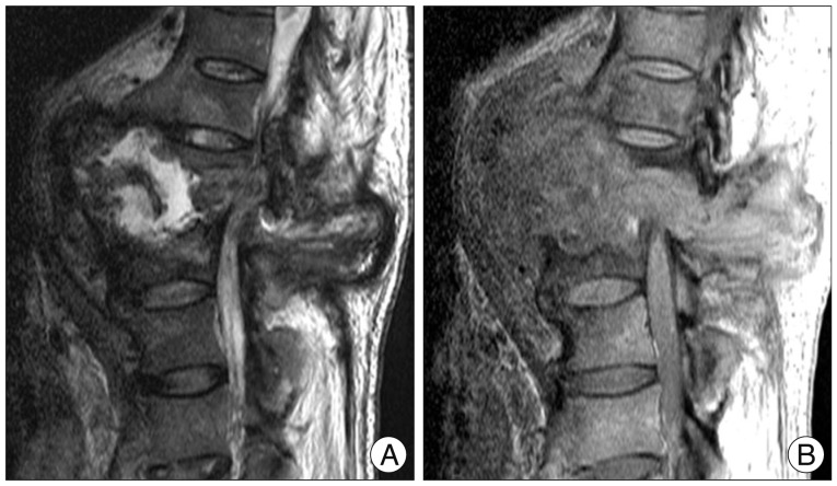
Fig. 3 Magnetic resonance imaging scans showing the complexity of the vertebral destruction and paraspinal mass with associated fluid collections. The mass is intermediate signal intensity on T1 weighted image (A), complex with mildly hyperintense areas on T2 weighted image (B).
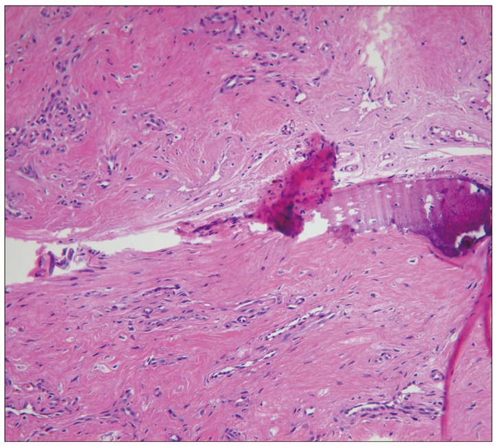
Fig. 4 Photomicrograph shows fibrous and bony tissue with hemosiderin pigment and hyalinization (H&E stain ×100).
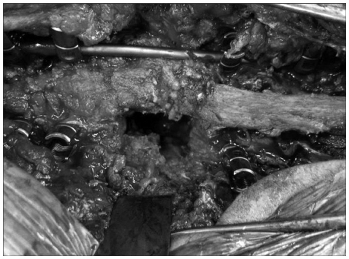
Fig. 5 Intraoperative photograph shows the posterolateral exposure of the Charcot segment, with a temporary stabilizing rod in place before distraction.
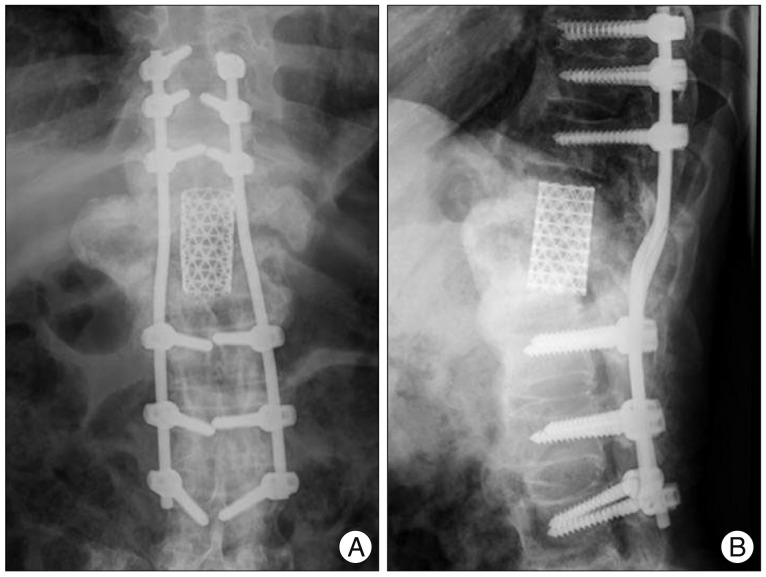
Fig. 6 The two years later follow up thoracolumbar sitting (A) AP and (B)lateral radiograph shows solid fusion and good alignment without evidence of hardware loosening.
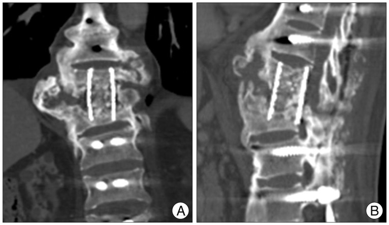
Fig. 7 Thoracolumbar (A) coronal and (B) sagittal CT image shows inadequate preparation of T10 lower and L1 upper end plate, and solid fusion between the residual T11, 12 bodies by a mesh cage.
Reference
-
1. Barrey C, Massourides H, Cotton F, Perrin G, Rode G. Charcot spine : two new case reports and a systematic review of 109 clinical cases from the literature. Ann Phys Rehabil Med. 2010; 53:200–220. PMID: 20338837.
Article2. Choi DY, Kim YB, Yeo SJ, Park SW, Hwang SN. One stage spondylodesis for the bursting fracture of the thoracolumbar spine through unilateral posterior approach. J Korean Neurosurg Soc. 2004; 35:379–382.3. Crim JR, Bassett LW, Gold RH, Mirra JM, Mikulics M, Dawson EG, et al. Spinal neuroarthropathy after traumatic paraplegia. AJNR Am J Neuroradiol. 1988; 9:359–362. PMID: 3128083.4. David KS, Agarwala AO, Rampersaud YR. Charcot arthropathy of the lumbar spine treated using one-staged posterior three-column shortening and fusion. Spine (Phila Pa 1976). 2010; 35:E657–E662. PMID: 20505559.
Article5. Devlin VJ, Ogilvie JW, Transfeldt EE, Boachie-Adjei O, Bradford DS. Surgical treatment of neuropathic spinal arthropathy. J Spinal Disord. 1991; 4:319–328. PMID: 1802163.
Article6. Eloesser L. On the nature of neufopathic affections of the joints. Ann Surg. 1917; 66:201–207. PMID: 17863764.7. Gupta R. A short history of neuropathic arthropathy. Clin Orthop Relat Res. 1993; (296):43–49. PMID: 8222448.
Article8. Harrison MJ, Sacher M, Rosenblum BR, Rothman AS. Spinal Charcot arthropathy. Neurosurgery. 1991; 28:273–277. PMID: 1997897.
Article9. Haus BM, Hsu AR, Yim ES, Meter JJ, Rinsky LA. Long-term follow-up of the surgical management of neuropathic arthropathy of the spine. Spine J. 2010; 10:e6–e16. PMID: 20494808.
Article10. Hoppenfeld S, Gross M, Giangarra C. Nonoperative treatment of neuropathic spinal arthropathy. Spine (Phila Pa 1976). 1990; 15:54–56. PMID: 2326701.
Article11. Jacobs WB, Bransford RJ, Bellabarba C, Chapman JR. Surgical management of Charcot spinal arthropathy : a single-center retrospective series highlighting the evolution of management. J Neurosurg Spine. 2012; 17:422–431. PMID: 22938550.
Article12. Kim YS, Cho YE. One stage three column fixation by posterior approach in thoracolumbar junction lesion. J Korean Neurosurg Soc. 1998; 27:222–228.13. Morita M, Miyauchi A, Okuda S, Oda T, Yamamoto T, Iwasaki M. Charcot spinal disease after spinal cord injury. J Neurosurg Spine. 2008; 9:419–426. PMID: 18976172.
Article14. Park YH, Taylor JA, Szollar SM, Resnick D. Imaging findings in spinal neuroarthropathy. Spine (Phila Pa 1976). 1994; 19:1499–1504. PMID: 7939982.
Article15. Rose DM, Hilton AI, Tucker SK. Charcot spinal arthropathy in a paraplegic weight lifter : case report. Spine (Phila Pa 1976). 2006; 31:E339–E341. PMID: 16688025.17. Standaert C, Cardenas DD, Anderson P. Charcot spine as a late complication of traumatic spinal cord injury. Arch Phys Med Rehabil. 1997; 78:221–225. PMID: 9041906.
Article18. Suda Y, Saito M, Shioda M, Kato H, Shibasaki K. Infected Charcot spine. Spinal Cord. 2005; 43:256–259. PMID: 15672097.
Article19. Vialle R, Mary P, Tassin JL, Parker F, Guillaumat M. Charcot's disease of the spine : diagnosis and treatment. Spine (Phila Pa 1976). 2005; 30:E315–E322. PMID: 15928542.
- Full Text Links
-
- Actions
-
Cited
- CITED
-
- Close
- Share
-
- Similar articles
-
- Charcot Arthropathy of the Lumbosacral Spine Mimicking a Vertebral Tumor after Spinal Cord Injury
- Unilateral Paramedian Transpedicular Approach for Repair of Anterior Transdural Spinal Cord Herniation: Report of a Case and Literature Review
- Posterolateral Approach for Ventral or Ventrolateral Thoracolumbar Lesion
- Clinical Experience of Traumatic C7-T1 Spondyloptosis
- Posterolateral Approach in Ventral or Ventrolateral Thoracic Tumor
