Anatomic Consideration of the C1 Laminar Arch for Lateral Mass Screw Fixation via C1 Lateral Lamina : A Landmark between the Lateral and Posterior Lamina of the C1
- Affiliations
-
- 1Department of Neurosurgery, Gangneung Asan Hospital, University of Ulsan College of Medicine, Gangneung, Korea. moonkyu71@empal.com
- 2Department of Anatomy, Catholic Institute for Applied Anatomy, College of Medicine, The Catholic University of Korea, Seoul, Korea.
- 3Department of Anatomy, College of Medicine, Chung-Ang University, Seoul, Korea.
- 4Department of Neurosurgery, Donghae Dong-In Hospital, Donghae, Korea.
- KMID: 2190853
- DOI: http://doi.org/10.3340/jkns.2013.54.1.25
Abstract
OBJECTIVE
To clarify the landmark for deciding the entry point for C1 lateral mass screws via the posterior arch by using 3-dimensional (3D) computed images.
METHODS
Resnick insisted that the C1 posterior arch could be divided into pure posterior and lateral lamina (C1 pedicle). Authors studied where this transition point (TP) is located between the posterior lamina and the C1 pedicle and how it can be recognized. The 3D computed images of 86 cadaver C1s (M : F=45 : 41) were used in this study.
RESULTS
The superior ridge of the C1 posterior arch had 2 types of orientation. One was in the vertical direction in the C1 posterior lamina and the other was in the horizontal direction in the C1 pedicle. The TP was located at the border between the 2 areas, the same site as the posterior end of the groove of the vertebral artery. On posterior-anterior projection, the posterior arch was sharpened abruptly at TP. We were unable to identify the TP in 6.4% of specimens due to complete or partial osseous bridges. A total of 93.8% of the TP were located between the most enlarged point of the spinal canal and the medial wall of the vertebral artery.
CONCLUSION
The anatomic entry zone of C1 lateral laminar screws was clarified and identified based on the TP by using preoperative 3D computed images.
MeSH Terms
Figure
-

Fig. 1 Definition of the transition point (TP). A : The posterior end (asterisk) of the groove of the vertebral artery can be seen in the posterior view from slightly above the lamina by 10-15°. B : The same site (arrow head) is denoted in the superior view of the atlas. C-F : Images are posterior views of C1 in various angles (C : +30°, D : +15°, E : 0°, F : -30°, asterisk : TP).
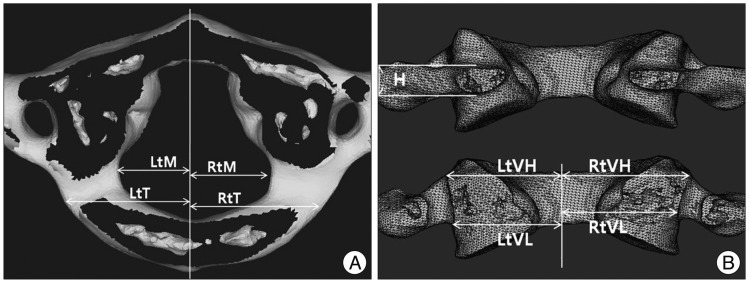
Fig. 2 Two orthogonal views (A : top; B : posterior side) of an atlas. H : minimal height of the C1 lateral lamina (C1 pedicle), Lt/Rt : left and right, M : maximal spinal canal diameter from midline, T : distance from midline to TP, VH : distance from midline to the medial side of the transverse foramen outlet, VL : distance from midline to the medial side of the transverse foramen inlet.

Fig. 3 Relative laterality ratio of a TP. This shows where a transition point is located comparing the safety zone between the spinal canal and the transverse foramen. Ratio=T/(VL - M). TP : transition point, VL : distance from midline to the medial side of the transverse foramen inlet, T : distance from midline to TP, M : maximal spinal canal diameter from midline.

Fig. 4 Lateral margin of the atlanto-occipital membrane (arrow heads).
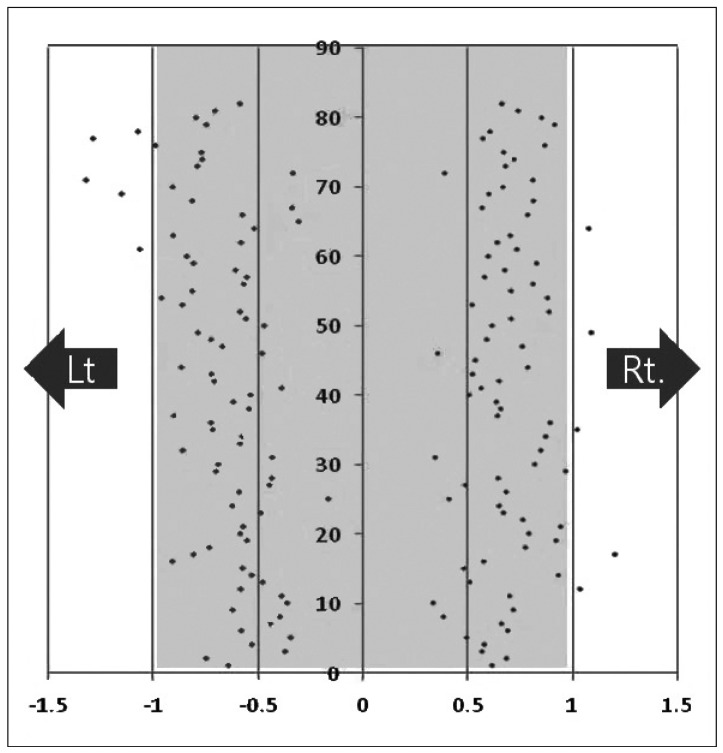
Fig. 5 Distribution of TPs. Individual dots show the relative laterality ratio of TP. X-axis : the relative laterality ratio, Y-axis : cadaver number, TP : transition point.
Cited by 1 articles
-
The Clinical Experience of Computed Tomographic-Guided Navigation System in C1-2 Spine Instrumentation Surgery
Sang-Uk Kim, Byoung-il Roh, Seong-Joon Kim, Sang-Don Kim
J Korean Neurosurg Soc. 2014;56(4):330-333. doi: 10.3340/jkns.2014.56.4.330.
Reference
-
1. Christensen DM, Eastlack RK, Lynch JJ, Yaszemski MJ, Currier BL. C1 anatomy and dimensions relative to lateral mass screw placement. Spine (Phila Pa 1976). 2007; 32:844–848. PMID: 17426627.
Article2. Chun HJ, Bak KH. Targeting a safe entry point for c2 pedicle screw fixation in patients with atlantoaxial instability. J Korean Neurosurg Soc. 2011; 49:351–354. PMID: 21887393.
Article3. Gebauer M, Barvencik F, Briem D, Kolb JP, Seitz S, Rueger JM, et al. Evaluation of anatomic landmarks and safe zones for screw placement in the atlas via the posterior arch. Eur Spine J. 2010; 19:85–90. PMID: 19882180.
Article4. Goel A, Kulkarni AG. Re : Tan M, Wang H, Wang Y, et al. : Morphometric evaluation of screw fixation in atlas via posterior arch and lateral mass. Spine. 2003;28:888-95. Spine (Phila Pa 1976). 2004; 29:1706. author reply 1706. PMID: 15284522.5. Goel A, Laheri V. Plate and screw fixation for atlanto-axial subluxation. Acta Neurochir (Wien). 1994; 129:47–53. PMID: 7998495.
Article6. Harms J, Melcher RP. Posterior C1-C2 fusion with polyaxial screw and rod fixation. Spine (Phila Pa 1976). 2001; 26:2467–2471. PMID: 11707712.
Article7. Karau PB, Ogengo JA, Hassanali J, Odula P. Anatomy and prevalence of atlas vertebrae bridges in a Kenyan population : an osteological study. Clin Anat. 2010; 23:649–653. PMID: 20533509.
Article8. Kobayashi Y, Kikuchi S, Konno S, Sekiguchi M. Insertion of lateral mass screw of the atlas via the posterior arch : anatomical study of screw insertion using dry bone samples of the atlas from Japanese cadavers. J Orthop Sci. 2008; 13:452–455. PMID: 18843460.
Article9. Lamberty BG, Zivanović S. The retro-articular vertebral artery ring of the atlas and its significance. Acta Anat (Basel). 1973; 85:113–122. PMID: 4197316.
Article10. Lee MJ, Cassinelli E, Riew KD. The feasibility of inserting atlas lateral mass screws via the posterior arch. Spine (Phila Pa 1976). 2006; 31:2798–2801. PMID: 17108832.
Article11. Ma XY, Yin QS, Wu ZH, Xia H, Liu JF, Xiang M, et al. C1 pedicle screws versus C1 lateral mass screws : comparisons of pullout strengths and biomechanical stabilities. Spine (Phila Pa 1976). 2009; 34:371–377. PMID: 19214096.12. Ma XY, Yin QS, Wu ZH, Xia H, Liu JF, Zhong SZ. Anatomic considerations for the pedicle screw placement in the first cervical vertebra. Spine (Phila Pa 1976). 2005; 30:1519–1523. PMID: 15990666.
Article13. Ma XY, Yin QS, Wu ZH, Xia H, Zhong SZ, Liu JF, et al. [Anatomic identification of the location of the pedicle of atlas with the lateral mass of C2 to C4 as the landmark]. Zhonghua Wai Ke Za Zhi. 2005; 43:774–776. PMID: 16083577.14. Murakami S, Mizutani J, Fukuoka M, Kato K, Sekiya I, Okamoto H, et al. Relationship between screw trajectory of C1 lateral mass screw and internal carotid artery. Spine (Phila Pa 1976). 2008; 33:2581–2585. PMID: 19011539.
Article15. Pan J, Li L, Qian L, Tan J, Sun G, Li X. C1 lateral mass screw insertion with protection of C1-C2 venous sinus : technical note and review of the literature. Spine (Phila Pa 1976). 2010; 35:E1133–E1136. PMID: 20885280.16. Resnick DK, Benzel EC. C1-C2 pedicle screw fixation with rigid cantilever beam construct : case report and technical note. Neurosurgery. 2002; 50:426–428. PMID: 11844283.
Article17. Resnick DK, Lapsiwala S, Trost GR. Anatomic suitability of the C1-C2 complex for pedicle screw fixation. Spine (Phila Pa 1976). 2002; 27:1494–1498. PMID: 12131706.
Article18. Rhee WT, You SH, Kim SK, Lee SY. Troublesome occipital neuralgia developed by c1-c2 harms construct. J Korean Neurosurg Soc. 2008; 43:111–113. PMID: 19096615.
Article19. Rocha R, Safavi-Abbasi S, Reis C, Theodore N, Bambakidis N, de Oliveira E, et al. Working area, safety zones, and angles of approach for posterior C-1 lateral mass screw placement : a quantitative anatomical and morphometric evaluation. J Neurosurg Spine. 2007; 6:247–254. PMID: 17355024.
Article20. Simsek S, Yigitkanli K, Comert A, Acar HI, Seckin H, Er U, et al. Posterior osseous bridging of C1. J Clin Neurosci. 2008; 15:686–688. PMID: 18378457.
Article21. Tan M, Wang H, Wang Y, Zhang G, Yi P, Li Z, et al. Morphometric evaluation of screw fixation in atlas via posterior arch and lateral mass. Spine (Phila Pa 1976). 2003; 28:888–895. PMID: 12942004.
Article22. Williams PL, Warwick R, Dyson M, Bannister LH. Gray's Anatomy. ed 37. New York: Churchill Livingstone, Inc;1989. p. 494.
- Full Text Links
-
- Actions
-
Cited
- CITED
-
- Close
- Share
-
- Similar articles
-
- Atlantoaxial Stabilization Using C1 Lateral Mass and C2 Pedicle/Translaminar Screw Fixation by Intraoperative C1- and C2-Direct-Captured Navigation with Preoperative Computed Tomography Images
- The Intersection Between Lateral Mass and Inferomedial Edge of the C1 Posterior Arch: A Reference Point for C1 Lateral Mass Screw Insertion
- Posterior Atlantoaxial Fixation with Lateral Mass Screw in the Atlas and Pedicle Screw in the Axis
- Old Atlantoaxial Rotary Subluxation Associated with High-riding Vertebral Arteries: Arthrodesis Using C1 Lateral Mass Screws and C2 Laminar Screws: A Case Report
- Unusual Anterior Arch Fracture of C1
