J Korean Neurosurg Soc.
2013 Mar;53(3):155-160. 10.3340/jkns.2013.53.3.155.
Risk Factors for Developing Large Emboli Following Carotid Artery Stenting
- Affiliations
-
- 1Department of Neurosurgery, Hanyang University Guri Hospital, Guri, Korea. cjh2324@hanyang.ac.kr
- 2Department of Radiology, Hanyang University Guri Hospital, Guri, Korea.
- KMID: 2190745
- DOI: http://doi.org/10.3340/jkns.2013.53.3.155
Abstract
OBJECTIVE
The introduction and development of the embolic protecting device (EPD) has resulted in a decreased rate of stroke after carotid artery stenting (CAS). The authors performed a retrospective study to investigate the risk factors for developing large emboli after CAS which can lead to ischemic events.
METHODS
A total of 35 consecutive patients who underwent CAS between January 2009 and March 2012 were included in this study. Patients were divided into two groups including those with small emboli (group A; grade 1, 2) and those with large emboli (group B; grade 3, 4). The size and number of emboli were assigned one of four grades (1=no clots, 2=1 or 2 small clots, 3=more than 3 small clots, 4=large clots) by microscopic observation of the EPD after CAS. We compared demographic characteristics, medical history, and angiographic findings of each group.
RESULTS
Thirty-five patients underwent CAS, and technical success was achieved in all cases. Twenty-three patients were included in group A and 12 patients in group B. Our results demonstrated that advanced age [odds ratio (OR) 1.24; 95% confidence interval (CI) 1.01-1.52; p=0.044] and smoking (OR 42.06; CI 2.828-625.65, p=0.006) were independent risk factors for developing large emboli after CAS.
CONCLUSION
In patients with carotid artery stenosis treated with CAS, advanced age and smoking increased the number and size of emboli. Although use of an EPD is controversial, it may be useful in CAS in patients with risk factors for large emboli in order to reduce the risk of ischemic events.
MeSH Terms
Figure
-

Fig. 1 Lateral carotid angle is measured by angle between A and B.
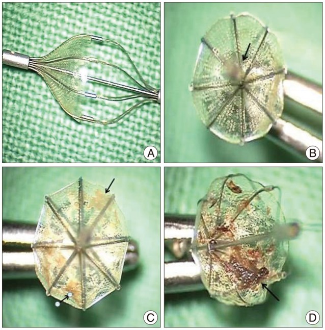
Fig. 2 Microscopic findings of the embolic protection devices after procedure. Arrows indicate the emboli captured. A : Grade 1. No emboli are visible. B : Grade 2. One small embolus is seen. C : Grade 3. Many clots not exceeding 3 mm are seen. D : Grade 4. Large clots over 3 mm are captured by the embolic protection device.
Cited by 1 articles
-
Findings of Angiography and Carotid Vessel Wall Imaging Associated with Post-Procedural Clinical Events after Carotid Artery Stenting
Sujin Jeon, Heejae Park, Hyo Sung Kwak, Seung Bae Hwang
Neurointervention. 2024;19(1):14-23. doi: 10.5469/neuroint.2023.00486.
Reference
-
1. Bacharach JM, Slovut DP, Ricotta J, Sullivan TM. Octogenarians are not at increased risk for periprocedural stroke following carotid artery stenting. Ann Vasc Surg. 2010; 24:153–159. PMID: 19748765.
Article2. Baldi S, Zander T, Rabellino M, González G, Maynar M. Carotid artery stenting without angioplasty and cerebral protection : a single-center experience with up to 7 years' follow-up. AJNR Am J Neuroradiol. 2011; 32:759–763. PMID: 21349967.
Article3. Bendszus M, Koltzenburg M, Burger R, Warmuth-Metz M, Hofmann E, Solymosi L. Silent embolism in diagnostic cerebral angiography and neurointerventional procedures : a prospective study. Lancet. 1999; 354:1594–1597. PMID: 10560674.
Article4. Cardaioli P, Giordan M, Panfili M, Chioin R. Complication with an embolic protection device during carotid angioplasty. Catheter Cardiovasc Interv. 2004; 62:234–236. PMID: 15170718.
Article5. Castriota F, Cremonesi A, Manetti R, Liso A, Oshola K, Ricci E, et al. Impact of cerebral protection devices on early outcome of carotid stenting. J Endovasc Ther. 2002; 9:786–792. PMID: 12546579.
Article6. Chung J, Shin YS, Lim YC, Park SK. The clinical outcomes of 75 consecutive patients with cervical carotid artery stenosis treated by carotid artery stenting. J Korean Neurosurg Soc. 2009; 45:350–354. PMID: 19609418.
Article7. Diethrich EB, Ndiaye M, Reid DB. Stenting in the carotid artery : initial experience in 110 patients. J Endovasc Surg. 1996; 3:42–62. PMID: 8798126.8. Dumont TM, Rughani AI. National trends in carotid artery revascularization surgery. J Neurosurg. 2012; 116:1251–1257. PMID: 22482791.
Article9. Gröschel K, Ernemann U, Schnaudigel S, Wasser K, Nägele T, Kastrup A. A risk score to predict ischemic lesions after protected carotid artery stenting. J Neurol Sci. 2008; 273:112–115. PMID: 18692206.
Article10. Gurm HS, Yadav JS, Fayad P, Katzen BT, Mishkel GJ, Bajwa TK, et al. Long-term results of carotid stenting versus endarterectomy in high-risk patients. N Engl J Med. 2008; 358:1572–1579. PMID: 18403765.
Article11. Kastrup A, Gröschel K, Krapf H, Brehm BR, Dichgans J, Schulz JB. Early outcome of carotid angioplasty and stenting with and without cerebral protection devices : a systematic review of the literature. Stroke. 2003; 34:813–819. PMID: 12624315.
Article12. Kastrup A, Gröschel K, Schnaudigel S, Nägele T, Schmidt F, Ernemann U. Target lesion ulceration and arch calcification are associated with increased incidence of carotid stenting-associated ischemic lesions in octogenarians. J Vasc Surg. 2008; 47:88–95. PMID: 18178458.
Article13. Lian X, Liu W, Li M, Lin M, Zhu S, Sun W, et al. Risk factors and complications associated with difficult retrieval of embolic protection devices in carotid artery stenting. Cardiovasc Intervent Radiol. 2012; 35:43–48. PMID: 21387123.
Article14. McDonnell CO, Fearn SJ, Baker SR, Goodman MA, Price D, Lawrence-Brown MM. Value of diffusion-weighted MRI during carotid angioplasty and stenting. Eur J Vasc Endovasc Surg. 2006; 32:46–50. PMID: 16546413.
Article15. Sayeed S, Stanziale SF, Wholey MH, Makaroun MS. Angiographic lesion characteristics can predict adverse outcomes after carotid artery stenting. J Vasc Surg. 2008; 47:81–87. PMID: 18178457.
Article16. Serrador JM, Picot PA, Rutt BK, Shoemaker JK, Bondar RL. MRI measures of middle cerebral artery diameter in conscious humans during simulated orthostasis. Stroke. 2000; 31:1672–1678. PMID: 10884472.
Article17. Tedesco MM, Coogan SM, Dalman RL, Haukoos JS, Lane B, Loh C, et al. Risk factors for developing postprocedural microemboli following carotid interventions. J Endovasc Ther. 2007; 14:561–567. PMID: 17696633.
Article18. Tedesco MM, Lee JT, Dalman RL, Lane B, Loh C, Haukoos JS, et al. Postprocedural microembolic events following carotid surgery and carotid angioplasty and stenting. J Vasc Surg. 2007; 46:244–250. PMID: 17600657.
Article19. Veith FJ, Amor M, Ohki T, Beebe HG, Bell PR, Bolia A, et al. Current status of carotid bifurcation angioplasty and stenting based on a consensus of opinion leaders. J Vasc Surg. 2001; 33(2 Suppl):S111–S116. PMID: 11174821.
Article20. Wu YM, Wong HF, Chen YL, Wong MC, Toh CH. Carotid stenting of asymptomatic and symptomatic carotid artery stenoses with and without the use of a distal embolic protection device. Acta Cardiol. 2011; 66:453–458. PMID: 21894801.
Article21. Yadav JS, Wholey MH, Kuntz RE, Fayad P, Katzen BT, Mishkel GJ, et al. Protected carotid-artery stenting versus endarterectomy in high-risk patients. N Engl J Med. 2004; 351:1493–1501. PMID: 15470212.
Article22. Zarins CK. Carotid endarterectomy : the gold standard. J Endovasc Surg. 1996; 3:10–15. PMID: 8798120.
- Full Text Links
-
- Actions
-
Cited
- CITED
-
- Close
- Share
-
- Similar articles
-
- A Case of Transseptal Approach to Carotid Artery Stenting in Right Internal Carotid Stenosis
- Carotid Artery Stenting
- Carotid Artery Stenting in Patients with Critical Stenosis of Proximal Internal Carotid Artery and Large Distal Arterial Thrombus: 2 Case Reports
- Carotid Artery Stenting for Asymptomatic Carotid Stenosis: What We Need to Know for Treatment Decision
- Safety of Carotid Artery Stenting in Elderly Patients with Severe Carotid Artery Stenosis
