Spinal Epidural Arteriovenous Hemangioma Mimicking Lumbar Disc Herniation
- Affiliations
-
- 1Department of Neurosurgery, Seoul National University Hospital, Seoul, Korea.
- 2Department of Neurosurgery, Seoul National University, Boramae Medical Center, Seoul, Korea. nslee@snu.ac.kr
- 3Department of Neurosurgery, Seoul National University College of Medicine, Seoul, Korea.
- KMID: 2190661
- DOI: http://doi.org/10.3340/jkns.2012.52.4.407
Abstract
- A spinal epidural hemangioma is rare. In this case, a 51 year-old female patient had low back pain and right thigh numbness. She was initially misdiagnosed as having a ruptured disc with possible sequestration of granulation tissue formation due to the limited number of spinal epidural hemangiomas and little-known radiological findings. Because there are no effective diagnostic tools to verify the hemangioma, more effort should be put into preoperative imaging tests to avoid misdiagnosis and poor decisions).
Keyword
Figure
-

Fig. 1 A spine MR image (at present). A and D : T2 wighted image. B and E : T1 weighted image. C and F : T1 enhanced image. The figures show a L3 level posterior epidural space ovoid shape enhancing lesion with heterogeneous T2 high/low SI and T1 iso to low signal intensity.
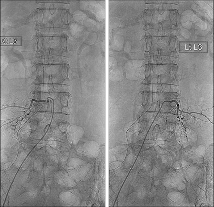
Fig. 2 A spinal angiography reveals no vascular abnormality.
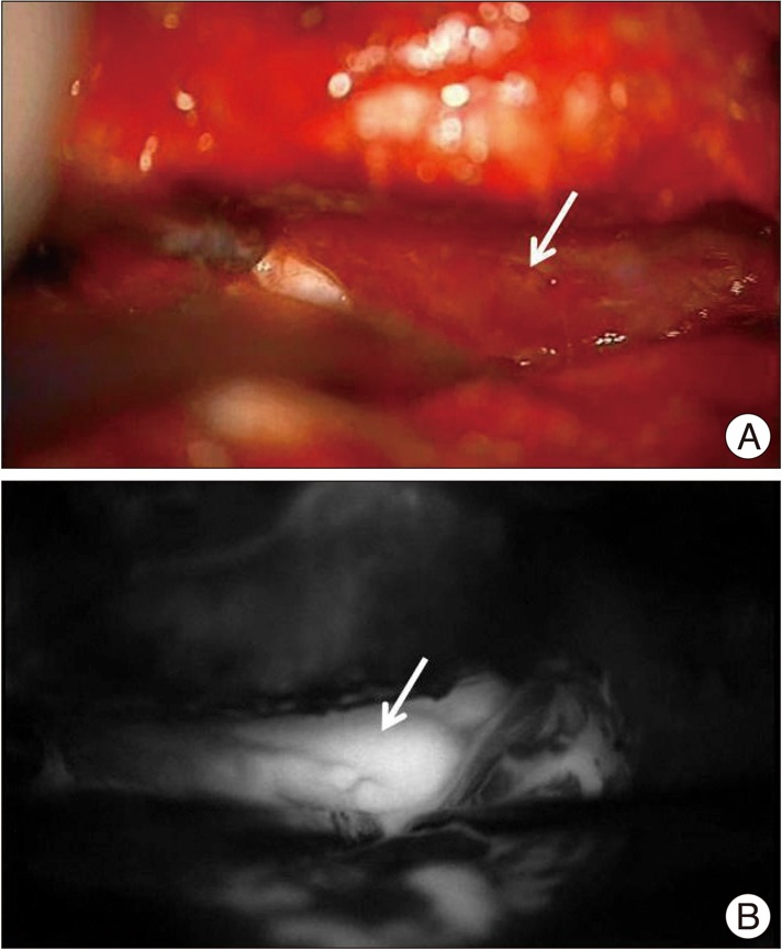
Fig. 3 A : The hyperemic vascular mass beneath the thecal sac. B : An ICG videoangiography shows delayed filling into the mass. ICG : indocyanine green.

Fig. 4 Histologically, the lesion reveals several anastomosing venous structures with irregular wall thickness (hematoxylin eosin ×40). A Masson's trichrome stain highlights abnormally thickened veins (inset) (×40).
Cited by 1 articles
-
Lumbar Epidural Varix Mimicking Disc Herniation
Adem Bursalı, Goktug Akyoldas, Ahmet Burak Guvenal, Onur Yaman
J Korean Neurosurg Soc. 2016;59(4):410-413. doi: 10.3340/jkns.2016.59.4.410.
Reference
-
1. Aoyagi N, Kojima K, Kasai H. Review of spinal epidural cavernous hemangioma. Neurol Med Chir (Tokyo). 2003; 43:471–475. discussion 476. PMID: 14620197.
Article2. Caruso G, Galarza M, Borghesi I, Pozzati E, Vitale M. Acute presentation of spinal epidural cavernous angiomas : case report. Neurosurgery. 2007; 60:E575–E576. discussion E576. PMID: 17327768.3. Colby GP, Coon AL, Sciubba DM, Bydon A, Gailloud P, Tamargo RJ. Intraoperative indocyanine green angiography for obliteration of a spinal dural arteriovenous fistula. J Neurosurg Spine. 2009; 11:705–709. PMID: 19951023.
Article4. Feider HK, Yuille DL. An epidural cavernous hemangioma of the spine. AJNR Am J Neuroradiol. 1991; 12:243–244. PMID: 1902020.5. Graziani N, Bouillot P, Figarella-Branger D, Dufour H, Peragut JC, Grisoli F. Cavernous angiomas and arteriovenous malformations of the spinal epidural space : report of 11 cases. Neurosurgery. 1994; 35:856–863. discussion 863-864. PMID: 7838334.6. Hong SP, Cho DS, Kim MH, Shin KM. Spinal epidural cavernous hemangioma simulating a disc protrusion : a case report. J Korean Neurosurg Soc. 2003; 33:509–511.7. Jo BJ, Lee SH, Chung SE, Paeng SS, Kim HS, Yoon SW, et al. Pure epidural cavernous hemangioma of the cervical spine that presented with an acute sensory deficit caused by hemorrhage. Yonsei Med J. 2006; 47:877–880. PMID: 17191320.
Article8. Lee JW, Cho EY, Hong SH, Chung HW, Kim JH, Chang KH, et al. Spinal epidural hemangiomas : various types of MR imaging features with histopathologic correlation. AJNR Am J Neuroradiol. 2007; 28:1242–1248. PMID: 17698523.
Article9. Minh NH. Cervicothoracic spinal epidural cavernous hemangioma : case report and review of the literature. Surg Neurol. 2005; 64:83–85. discussion 85. PMID: 15993196.
Article10. Raabe A, Beck J, Gerlach R, Zimmermann M, Seifert V. Near-infrared indocyanine green video angiography : a new method for intraoperative assessment of vascular flow. Neurosurgery. 2003; 52:132–139. discussion 139. PMID: 12493110.
Article11. Raabe A, Nakaji P, Beck J, Kim LJ, Hsu FP, Kamerman JD, et al. Prospective evaluation of surgical microscope-integrated intraoperative near-infrared indocyanine green videoangiography during aneurysm surgery. J Neurosurg. 2005; 103:982–989. PMID: 16381184.
Article12. Shin JH, Lee HK, Rhim SC, Park SH, Choi CG, Suh DC. Spinal epidural cavernous hemangioma : MR findings. J Comput Assist Tomogr. 2001; 25:257–261. PMID: 11242225.
- Full Text Links
-
- Actions
-
Cited
- CITED
-
- Close
- Share
-
- Similar articles
-
- Spontaneous Spinal Epidural Hematoma Mimicking Lumbar Disc Herniation
- Lumbar Epidural Venography in the Diagnosis of Lumbar Disc Herniation
- Lumbar Extradural Arteriovenous Malformation Mimicking Intervertebral Disc Herniation in the Lumbar Spine: A Case Report
- Redundant Nerve Roots of Cauda Equina Mimicking Intradural Disc Herniation: A Case Report
- Endovascular Treatment of Spinal Dural and Epidural Arteriovenous Fistula as Complication of Lumbar Surgery
