J Korean Neurosurg Soc.
2012 Aug;52(2):126-132. 10.3340/jkns.2012.52.2.126.
Imaging Findings of Solitary Spinal Bony Lesions and the Differential Diagnosis of Benign and Malignant Lesions
- Affiliations
-
- 1Department of Neurosurgery, Pusan National University Hospital, Busan, Korea. farlateral@hanmail.net
- 2Department of Radiology, Pusan National University Hospital, Busan, Korea.
- 3Department of Orthopedic Surgery, Medical Research Institute, Pusan National University Hospital, Busan, Korea.
- KMID: 2190551
- DOI: http://doi.org/10.3340/jkns.2012.52.2.126
Abstract
OBJECTIVE
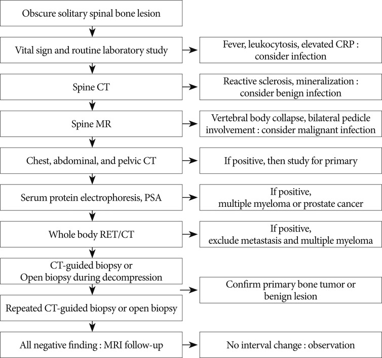
The purpose of this study was to present the MRI and CT findings of solitary spinal bone lesions (SSBLs) with the aims of aiding the differential diagnoses of malignant tumors and benign lesions, and proposing a diagnostic strategy for obscure SSBLs.
METHODS
The authors retrospectively reviewed the imaging findings of 19 patients with an obscure SSBL on MRI at our hospital from January 1994 to April 2011. The 19 patients were divided to benign groups and malignant groups according to final diagnosis. MRI and CT findings were evaluated and the results of additional work-up studies were conducted to achieve a differential diagnosis.
RESULTS
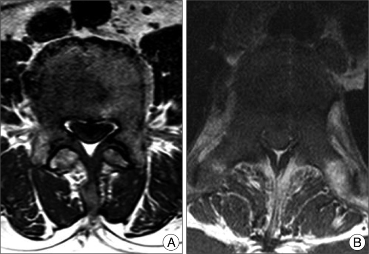
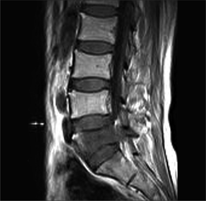
At final diagnoses, 10 (52.6%) of the 19 SSBLs were malignant tumors and 9 (47.4%) were benign lesions. The malignant tumors included 6 metastatic cancers, 3 multiple myelomas, and 1 chordoma, and the benign lesions included 4 osteomyelitis, 2 hemangiomas, 2 nonspecific chronic inflammations, and 1 giant cell tumor. No MRI characteristics examined was found to be significantly different in the benign and malignant groups. Reactive sclerotic change was observed by CT in 1 (10.0%) of the 10 malignant lesions and in 7 (77.8%) of the 9 benign lesions (p=0.005).
CONCLUSION
Approximately half of the obscure SSBLs were malignant tumors. CT and MRI findings in combination may aid the differential diagnosis of obscure SSBLs. In particular, sclerotic change on CT images was an important finding implying benign lesion. Finally, we suggest a possible diagnostic strategy for obscure SSBLs on MRI.
Keyword
MeSH Terms
Figure
Reference
-
1. Abdel Razek AA, Castillo M. Imaging appearance of primary bony tumors and pseudo-tumors of the spine. J Neuroradiol. 2010; 37:37–50. PMID: 19781780.
Article2. Aebi M. Spinal metastasis in the elderly. Eur Spine J. 2003; 12(Suppl 2):S202–S213. PMID: 14505120.
Article3. Destombe C, Botton E, Le Gal G, Roudaut A, Jousse-Joulin S, Devauchelle-Pensec V, et al. Investigations for bone metastasis from an unknown primary. Joint Bone Spine. 2007; 74:85–89. PMID: 17218141.
Article4. Dunbar JA, Sandoe JA, Rao AS, Crimmins DW, Baig W, Rankine JJ. The MRI appearances of early vertebral osteomyelitis and discitis. Clin Radiol. 2010; 65:974–981. PMID: 21070900.
Article5. Erlemann R. Imaging and differential diagnosis of primary bone tumors and tumor-like lesions of the spine. Eur J Radiol. 2006; 58:48–67. PMID: 16431065.
Article6. Gosfield E 3rd, Alavi A, Kneeland B. Comparison of radionuclide bone scans and magnetic resonance imaging in detecting spinal metastases. J Nucl Med. 1993; 34:2191–2198. PMID: 8254410.7. He MX, Zhu MH, Zhang YM, Fu QG, Wu LL. [Solitary plasmacytoma of spine : a clinical, radiologic and pathologic study of 13 cases]. Zhonghua Bing Li Xue Za Zhi. 2009; 38:307–311. PMID: 19575872.8. Hsu CY, Yu CW, Wu MZ, Chen BB, Huang KM, Shih TT. Unusual manifestations of vertebral osteomyelitis : intraosseous lesions mimicking metastases. AJNR Am J Neuroradiol. 2008; 29:1104–1110. PMID: 18356469.
Article9. Iizuka Y, Iizuka H, Tsutsumi S, Nakagawa Y, Nakajima T, Sorimachi Y, et al. Diagnosis of a previously unidentified primary site in patients with spinal metastasis : diagnostic usefulness of laboratory analysis, CT scanning and CT-guided biopsy. Eur Spine J. 2009; 18:1431–1435. PMID: 19533181.
Article10. Katagiri H, Takahashi M, Inagaki J, Sugiura H, Ito S, Iwata H. Determining the site of the primary cancer in patients with skeletal metastasis of unknown origin : a retrospective study. Cancer. 1999; 86:533–537. PMID: 10430264.
Article11. Min JW, Um SW, Yim JJ, Yoo CG, Han SK, Shim YS, et al. The role of whole-body FDG PET/CT, Tc 99m MDP bone scintigraphy, and serum alkaline phosphatase in detecting bone metastasis in patients with newly diagnosed lung cancer. J Korean Med Sci. 2009; 24:275–280. PMID: 19399270.
Article12. Puri A, Shingade VU, Agarwal MG, Anchan C, Juvekar S, Desai S, et al. CT-guided percutaneous core needle biopsy in deep seated musculoskeletal lesions : a prospective study of 128 cases. Skeletal Radiol. 2006; 35:138–143. PMID: 16391943.
Article13. Rimondi E, Rossi G, Bartalena T, Ciminari R, Alberghini M, Ruggieri P, et al. Percutaneous CT-guided biopsy of the musculoskeletal system : results of 2027 cases. Eur J Radiol. 2011; 77:34–42. PMID: 20832220.
Article14. Rodallec MH, Feydy A, Larousserie F, Anract P, Campagna R, Babinet A, et al. Diagnostic imaging of solitary tumors of the spine : what to do and say. Radiographics. 2008; 28:1019–1041. PMID: 18635627.
Article15. Samartzis D, Marco RA. Osteochondroma of the sacrum : a case report and review of the literature. Spine (Phila Pa 1976). 2006; 31:E425–E429. PMID: 16741444.16. Shih TT, Huang KM, Li YW. Solitary vertebral collapse : distinction between benign and malignant causes using MR patterns. J Magn Reson Imaging. 1999; 9:635–642. PMID: 10331758.
Article17. Skrzynski MC, Biermann JS, Montag A, Simon MA. Diagnostic accuracy and charge-savings of outpatient core needle biopsy compared with open biopsy of musculoskeletal tumors. J Bone Joint Surg Am. 1996; 78:644–649. PMID: 8642019.
Article18. Theodorou DJ, Theodorou SJ, Sartoris DJ. An imaging overview of primary tumors of the spine : Part 1. Benign tumors. Clin Imaging. 2008; 32:196–203. PMID: 18502347.
- Full Text Links
- Actions
-
Cited
- CITED
-
- Close
- Share
- Similar articles
-
- Imaging Findings of Spinal Metastases with Differential Diagnosis: Focusing on Solitary Spinal Lesion in Older Patients
- Solitary Pulmonary Nodule: CT Findings
- Destructive lesions of vertebral body:CT findings and differential diagnosis of inflammation and malignancy
- CT and MR Findings of Chest Wall Masses
- Differential Diagnosis of Vertebral Lesion by Magnetic Resonance Imaging


