J Korean Med Assoc.
2010 Jul;53(7):562-568. 10.5124/jkma.2010.53.7.562.
Imaging Diagnosis of Colorectal Cancer
- Affiliations
-
- 1Department of Radiology, University of Ulsan College of Medicine, Asan Medical Center, Seoul, Korea. aykim@amc.seoul.kr
- KMID: 2188330
- DOI: http://doi.org/10.5124/jkma.2010.53.7.562
Abstract
- Recently, treatment strategy of rectal cancer has undergone a dramatic change. Application of total mesorectal excision and preoperative chemoradiation therapy (PCRT) has become standard procedure for locoregional and locally advanced rectal cancer, respectively. Functional and morphological radiologic evaluation as well as multidisciplinary approach is both essential for planning patient-specific therapy. In other words, the needs for more accurate T-and N-staging and assessment of circumferential resection margin, both before and after PCRT, are increasing rapidly. Although there is no consensus on the role of diagnostic imaging such as endorectal ultrasonography, computed tomography and magnetic resonance imaging (MRI), in evaluation of rectal cancer patient so far, MRI is emerging as an essential imaging modality with superior trssue contrast and multiplanar approach.
Keyword
MeSH Terms
Figure
-

Figure 1 Magnetic resonance imaging (MRI) of rectal cancer. Arrows indicate the mesorectal fascia.
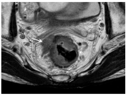
Figure 2 Magnetic resonance imaging (MRI) of rectal cancer. Arrow indicates positive circumferential resection margin by T3 rectal cancer.
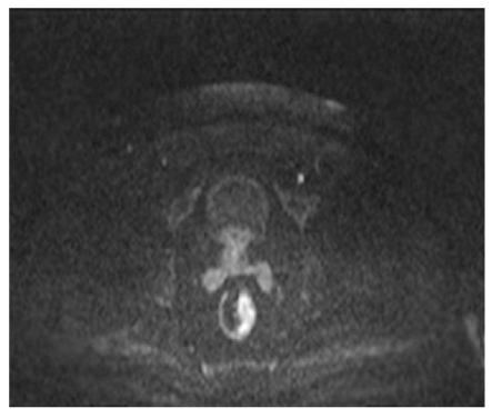
Figure 3 Diffusion-weighted imaging of rectal cancer. Viable cancer shows high signal intensity on diffusion-weighted image of rectal MRI.
Cited by 1 articles
-
An Understanding of New Concepts and Publication Ethics in the Use of Online Medical Journals and their Database Web Sites
Chong-Woo Bae, Chang-Kok Hahm
J Korean Med Assoc. 2010;53(8):685-694. doi: 10.5124/jkma.2010.53.8.685.
Reference
-
1. Heald RJ, Ryall RD. Recurrence and survival after total mesorectal excision for rectal cancer. Lancet. 1986. 28:1479–1482.
Article2. Barbaro B, Fiorucci C, Tebala C, Valentini V, Gambacorta MA, Vecchio FM, Rizzo G, Coco C, Crucitti A, Ratto C, Bonomo L. Locally Advanced Rectal Cancer: MR Imaging in Prediction of Response after Preoperative Chemotherapy and Radiation Therapy. Radiology. 2009. 250:730–739.
Article3. Brown G, Radcliffe AG, Newcombe RG, Dallimore NS, Bourne MW, Williams GT. Preoperative assessment of prognostic factors in rectal cancer using high-resolution magnetic resonance imaging. Br J Surg. 2003. 90:355–364.
Article4. Kapiteijn E, Marijnen CA, Nagtegaal ID, Putter H, Steup WH, Wiggers T, Rutten HJ, Pahlman L, Glimelius B, van Krieken JH, Leer JW, van de Velde CJ. Dutch Colorectal Cancer Group. Preoperative radiotherapy combined with total mesorectal excision for resectable rectal cancer. N Engl J Med. 2001. 345:638–646.
Article5. Colorectal Cancer Collaborative Group. Adjuvant radiotherapy for rectal cancer: a systematic overview of 8,507 patients from 22 randomised trials. Lancet. 2001. 358:1291–1304.6. Katsura Y, Yamada K, Ishizawa T, Yoshinaka H, Shimazu H. Endorectal ultrasonography for the assessment of wall invasion and lymph node metastasis in rectal cancer. Dis Colon Rectum. 1992. 35:362–368.
Article7. Garcia-Aguilar J, Pollack J, Lee SH, Hernandez de Anda E, Mellgren A, Wong WD, Finne CO, Rothenberger DA, Madoff RD. Accuracy of endorectal ultrasonography in preoperative staging of rectal tumors. Dis Colon Rectum. 2002. 45:10–15.
Article8. Gualdi GF, Casciani E, Guadalaxara A, d'Orta C, Polettini E, Pappalardo G. Local staging of rectal cancer with transrectal ultrasound and endorectal magnetic resonance imaging: comparison with histologic findings. Dis Colon Rectum. 2000. 43:338–345.
Article9. Akasu T, Kondo H, Moriya Y, Sugihara K, Gotoda T, Fujita S, Muto T, Kakizoe T. Endorectal ultrasonography and treatment of early stage rectal cancer. World J Surg. 2000. 24:1061–1068.
Article10. Thoeni RF, Moss AA, Schnyder P, Margulis AR. Detection and staging of primary rectal and rectosigmoid cancer by computed tomography. Radiology. 1981. 141:135–138.
Article11. Hodgman CG, MacCarty RL, Wolff BG, May GR, Berquist TH, Sheedy PF 2nd, Beart RW Jr, Spencer RJ. Preoperative staging of rectal carcinoma by computed tomography and 0.15T magnetic resonance imaging: preliminary report. Dis Colon Rectum. 1986. 29:446–450.
Article12. Zerhouni EA, Rutter C, Hamilton SR, Balfe DM, Megibow AJ, Francis IR, Moss AA, Heiken JP, Tempany CM, Aisen AM, Weinreb JC, Gatsonis C, McNeil BJ. CT and MR imaging in the staging of colorectal carcinoma: report of the Radiology Diagnostic Oncology Group II. Radiology. 1996. 200:443–451.
Article13. Thoeni RF. Colorectal cancer: radiologic staging. Radiol Clin North Am. 1997. 35:457–485.14. Chiesura-Corona M, Muzzio PC, Giust G, Zuliani M, Pucciarelli S, Toppan P. Rectal cancer: CT local staging with histopathologic correlation. Abdom Imaging. 2001. 26:134–138.
Article15. Hodgman CG, MacCarty RL, Wolff BG, May GR, Berquist TH, Sheedy PF 2nd, Beart RW Jr, Spencer RJ. Preoperative staging of rectal carcinoma by computed tomography and 0.15T magnetic resonance imaging: preliminary report. Dis Colon Rectum. 1986. 29:446–450.
Article16. Guinet C, Buy JN, Ghossain MA, Sézeur A, Mallet A, Bigot JM, Vadrot D, Ecoiffier J. Comparison of magnetic resonance imaging and computed tomography in the preoperative staging of rectal cancer. Arch Surg. 1990. 125:385–388.
Article17. Chan TW, Kressel HY, Milestone B, Tomachefski J, Schnall M, Rosato E, Daly J. Rectal carcinoma: staging at MR imaging with endorectal surface coil-work in progress. Radiology. 1991. 181:461–467.
Article18. Vogl TJ, Pegios W, Mack MG, Hünerbein M, Hintze R, Adler A, Lobbeck H, Hammerstingl R, Wust P, Schlag P, Felix R. Accuracy of staging rectal tumors with contrast-enhanced transrectal MR imaging. AJR Am J Roentgenol. 1997. 168:1427–1434.
Article19. Andreoni B, Chiappa A, Bertani E, Bellomi M, Orecchia R, Zampino M, Fazio N, Venturino M, Orsi F, Sonzogni A, Pace U, Monfardini L. Surgical outcomes for colon and rectal cancer over a decade: results from a consecutive monocentric experience in 902 unselected patients. World J Surg Oncol. 2007. 5:73.
Article20. Sagar PM, Pemberton JH. Surgical management of locally recurrent rectal cancer. Br J Surg. 1996. 83:293–304.
Article21. Quirke P, Durdey P, Dixon MF, Williams NS. Local recurrence of rectal adenocarcinoma due to inadequate surgical resection. Histopathological study of lateral tumour spread and surgical excision. Lancet. 1986. 2:996–999.22. Mönig SP, Baldus SE, Zirbes TK, Schröder W, Lindemann DG, Dienes HP, Hölscher AH. Lymph node size and metastatic infiltration in colon cancer. Ann Surg Oncol. 1999. 6:579–581.
Article23. Andreola S, Leo E, Belli F, Bufalino R, Tomasic G, Lavarino C, Baldini MT, Meroni E. Manual dissection of adenocarcinoma of the lower third of the rectum specimens for detection of lymph node metastases smaller than 5 mm. Cancer. 1996. 77:607–612.
Article24. Balthazar EJ, Megibow AJ, Hulnick D, Naidich DP. Carcinoma of the colon: detection and preoperative staging by CT. AJR Am J Roentgenol. 1988. 150:301–306.
Article25. Thompson WM, Halvorsen RA, Foster WL Jr, Roberts L, Gibbons R. Preoperative and postoperative CT staging of rectosigmoid carcinoma. AJR Am J Roentgenol. 1986. 146:703–710.
Article26. Holdsworth PJ, Johnston D, Chalmers AG, Chennells P, Dixon MF, Finan PJ, Primrose JN, Quirke P. Endoluminal ultrasound and computed tomography in the staging of rectal cancer. Br J Surg. 1988. 75:1019–1022.
Article27. Brown G, Richards CJ, Bourne MW, Newcombe RG, Radcliffe AG, Dallimore NS, Williams GT. Morphologic predictors of lymph node status in rectal cancer with use of highspatial-resolution MR imaging with histopathologic comparison. Radiology. 2003. 227:371–377.
Article28. Bellin MF, Roy C, Kinkel K, Thoumas D, Zaim S, Vanel D, Tuchmann C, Richard F, Jacqmin D, Delcourt A, Challier E, Lebret T, Cluzel P. Lymph node metastases: safety and effectiveness of MR imaging with ultrasmall superparamagnetic iron oxide particles-initial clinical experience. Radiology. 1998. 207:799–808.
Article29. Sigal R, Vogl T, Casselman J, Moulin G, Veillon F, Hermans R, Dubrulle F, Viala J, Bosq J, Mack M, Depondt M, Mattelaer C, Petit P, Champsaur P, Riehm S, Dadashitazehozi Y, de Jaegere T, Marchal G, Chevalier D, Lemaitre L, Kubiak C, Helmberger R, Halimi P. Lymph node metastases from head and neck squamous cell carcinoma: MR imaging with ultrasmall superparamagnetic iron oxide particles (Sinerem MR)-results of a phase-III multicenter clinical trial. Eur Radiol. 2002. 12:1104–1113.
Article30. Janjan NA, Khoo VS, Abbruzzese J, Pazdur R, Dubrow R, Cleary KR, Allen PK, Lynch PM, Glober G, Wolff R, Rich TA, Skibber J. Tumor downstaging and sphincter preservation with preoperative chemoradiation in locally advanced rectal cancer: the M. D. Anderson Cancer Center experience. Int J Radiat Oncol Biol Phys. 1999. 44:1027–1038.
Article31. Reerink O, Verschueren RC, Szabo BG, Hospers GA, Mulder NH. A favourable pathological stage after neoadjuvant radiochemotherapy in patients with initially irresectable rectal cancer correlates with a favourable prognosis. Eur J Cancer. 2003. 39:192–195.
Article32. Koh DM, Chau I, Tait D, Wotherspoon A, Cunningham D, Brown G. Evaluating mesorectal lymph nodes in rectal cancer before and after neoadjuvant chemoradiation using thin-section T2-weighted magnetic resonance imaging. Int J Radiat Oncol Biol Phys. 2008. 71:456–461.
Article33. Taylor FG, Swift RI, Blomqvist L, Brown G. A systematic approach to the interpretation of preoperative staging MRI for rectal cancer. AJR Am J Roentgenol. 2008. 191:1827–1835.
Article34. Rullier A, Laurent C, Capdepont M, Vendrely V, Bioulac-Sage P, Rullier E. Impact of tumor response on survival after radiochemotherapy in locally advanced rectal carcinoma. Am J Surg Pathol. 2010. 34:562–568.
Article35. Chen CC, Lee RC, Lin JK, Wang LW, Yang SH. How accurate is magnetic resonance imaging in restaging rectal cancer in patients receiving preoperative combined chemoradio-therapy? Dis Colon Rectum. 2005. 48:722–728.
Article36. Kuo LJ, Chern MC, Tsou MH, Liu MC, Jian JJ, Chen CM, Chung YL, Fang WT. Interpretation of magnetic resonance imaging for locally advanced rectal carcinoma after preoperative chemoradiation therapy. Dis Colon Rectum. 2005. 48:23–28.
Article37. Vliegen RF, Beets GL, Lammering G, Dresen RC, Rutten HJ, Kessels AG, Oei TK, de Bruïne AP, van Engelshoven JM, Beets-Tan RG. Mesorectal fascia invasion after neoadjuvant chemotherapy and radiation therapy for locally advanced rectal cancer: accuracy of MR imaging for prediction. Radiology. 2008. 246:454–462.
Article38. Allen SD, Padhani AR, Dzik-Jurasz AS, Glynne-Jones R. Rectal carcinoma: MRI with histologic correlation before and after chemoradiation therapy. AJR Am J Roentgenol. 2007. 188:442–451.
Article39. Hosonuma T, Tozaki M, Ichiba N, Sakuma T, Hayashi D, Yanaga K, Fukuda K. Clinical usefulness of diffusion-weighted imaging using low and high b-values to detect rectal cancer. Magn Reson Med Sci. 2006. 5:173–177.
Article40. Rao SX, Zeng MS, Chen CZ, Li RC, Zhang SJ, Xu JM, Hou YY. The value of diffusionweighted imaging in combination with T2-weighted imaging for rectal cancer detection. Eur J Radiol. 2008. 65:299–303.
Article41. DeVries AF, Kremser C, Hein PA, Griebel J, Krezcy A, Ofner D, Pfeiffer KP, Lukas P, Judmaier W. Tumor microcirculation and diffusion predict therapy outcome for primary rectal carcinoma. Int J Radiat Oncol Biol Phys. 2003. 56:958–965.
Article42. Hein PA, Kremser C, Judmaier W, Griebel J, Pfeiffer KP, Kreczy A, Hug EB, Lukas P, DeVries AF. Diffusion-weighted magnetic resonance imaging for monitoring diffusion changes in rectal carcinoma during combined, preoperative chemoradiation: preliminary results of a prospective study. Eur J Radiol. 2003. 45:214–222.43. Dzik-Jurasz A, Domenig C, George M, Wolber J, Padhani A, Brown G, Doran S. Diffusion MRI for prediction of response of rectal cancer to chemoradiation. Lancet. 2002. 360:307–308.
Article
- Full Text Links
-
- Actions
-
Cited
- CITED
-
- Close
- Share
-
- Similar articles
-
- Endoscopic diagnosis and treatment of early colorectal cancer
- Introduction: What Are New Roles of Current Colonoscopy?
- Clinical Applications of Linked Color Imaging and Blue Laser/Light Imaging in the Screening, Diagnosis, and Treatment of Superficial Colorectal Tumors
- Equipment-Based Image-Enhanced Endoscopy for Differentiating Colorectal Polyps
- Hereditary Nonpolyposis Colorectal Cancer
